“The Radiology Reset Button – overcoming the normalcy bias”

Fodi KyriakosFodi Kyriakos explores how the COVID-19 pandemic could be the catalyst for change in radiology and encourages our community to grasp the opportunity to “seize the moment”  and plan for recovery.

At the beginning of 2020, if someone had told radiology leaders that all NHS outstanding reporting backlogs would be reduced to virtually zero by May, I’m sure they would have looked at you in disbelief and asked what sorcery had been involved, but this situation is exactly where we find ourselves today.


Normalcy Bias – Noun [edit]

normalcy bias (plural normalcy biases)

The phenomenon of disbelieving one’s situation when faced with grave and imminent danger and/or catastrophe. As in over focusing on the actual phenomenon instead of taking evasive action, a state of paralysis.

Historical challenges

In the past, it has often taken lots of effort to either invoke or accept change of any kind in radiology and for those managing services, there’s also been a certain amount of risk associated with putting your head above the parapet or being a trailblazer. It has been sometimes easier to follow the well-trodden path rather than to create a new one. Workloads and budgetary constraints have also been a disabler, restricting decision making to the ‘here and now’. This has resulted in failing, or in most cases, not being able to foresee or plan for events that have never happened before, such as an event like a pandemic crisis. Psychology refers to this state of being as normalcy bias. For those who are not familiar with the term, you will certainly be aware of its connotations and radiology now finds itself at this cross-roads.

Ever since the introduction of digital radiography and PACS, NHS radiology reporting backlogs have been a contentious issue among experts, and a recurring feature in the mainstream media! Often being highlighted (and with some justification) in relation to areas such as missed cancer diagnosis, where even the slightest of delays can have a significant bearing on the overall outcome.

Serious backlogs

The extent to which backlogs were a serious issue in the UK was further exacerbated by various Care Quality Commission (CQC)  inspections, which raised concerns regarding reporting backlogs that resulted in delayed or missed diagnosis of conditions that may have otherwise been picked up.

By the end of February 2020, the situation of backlogs was as much an issue as at any time before. Insufficient reporting capacity had led to a build-up of outstanding reports, which in turn meant that outsourcing was at its highest ever levels and growing pressures to meet new deadlines, such as the cancer pathway targets, were increasingly exposing the lack of options available to resolve the problem.

So, you would have been excused if you thought that a crisis such as the COVID-19 pandemic would simply exacerbate the reporting challenges facing radiology. However, this has not been the case. Instead, we have witnessed radiology’s own “clear the decks” exercise, where in fact the complete opposite situation has occurred, resulting in backlogs across the UK being virtually eliminated. Who would have thought that the worst crisis to hit the country (and the world) in 75 years would be a catalyst for NHS radiology departments to press the reset button?

Reset image

Of course, we recognise the superficial nature of this situation. During the pandemic, practically all routine referral activity came to a grinding halt, which allowed radiology to concentrate on COVID-19 and Emergency Department (ED) patients. Chest X-rays and CTs were identified as two of the key diagnostic tools for the virus, but the volumes were manageable. Accident and Emergency footfall was reduced to almost 50% of its usual figures, so reporters were practically able to deliver a ‘Hot-Reporting’ examination for every patient requiring imaging. Something which ED and Intensive care unit (ICU) consultants have grown quickly accustomed to.

During this time, radiology was also still required to work to critical staffing levels, so radiographers and radiologists were covering 24/7 rotas, but due to the lack of activity outside of portable X-ray scanning in ICU, many staff were not being utilised. So, while this enabled the catch up in radiology reporting to take place, what we witnessed was the ‘ying and yang’ of radiology. On the one hand, integral to the continuity of a patient’s pathway and critical to defining an outcome – AND on the other hand, completely dependent on throughput from referrers to maintain activity levels.

Seizing the moment!

So what happens next? Well, in a world where we can guarantee almost nothing, in this situation, we can guarantee that radiology will remain the centre point for the recovery phase of the pandemic, but with the added challenge of complying to ‘social distancing’ and ‘equipment cleaning’ guidelines, how do we manage the continuation of treating COVID-19 patients, while reintroducing ‘business as usual’ and ‘deferred’ patients whose treatment has been delayed?

The “Reset Button” has enabled something else to happen. For the first time, there is now some headspace to plan for the recovery phase and for the next phase at least, there is now funding available to support the recovery. So how do we avoid going back to where we were before the pandemic? How do we seize the moment?

Time to make the changes!

Albert Einstein once famously said: “We can’t solve problems by using the same kind of thinking we used when we created them.” This quote has never been more poignant in the present day and while the pressure to manage change will be at its highest, this is the right time to make these changes happen! With the benefit of ‘The Reset Button’, if we can learn from the past and apply new ways of working moving forward, we can avoid falling into the trap of the normalcy bias and witness the radiology reset button offering a new, efficient and more streamlined radiology department moving forward.

Everything you wanted to know about radiology but were afraid to ask…

On Wednesday 17 June, a live event organised by InHealth, in partnership with The British Institute of Radiology and the Society of Radiographers is taking place, titled: “The Radiology reset button has been pressed”. The aim is to tackle these challenges and support radiology managers as they enter the recovery phase. It will bring together senior figures from radiology and within healthcare to offer insights, opinions and advice on how we can approach this coming period and use what positives we have experienced during the pandemic to create service improvements throughout radiology.

There will be opportunities for radiology managers, clinical leads, radiographers and radiologists to put their questions to the speakers in the panel discussions after their presentations.

REGISTER FOR THE RADIOLOGY RESET BUTTON HAS BEEN PRESSED HERE

(The event is free for all)
About Fodi Kyriakos

Mr Fodi Kyriakos is a former director of RIG Healthcare and founder of RIG Reporting,
the UK’s first provider of external radiographer reporting services. In 2016 he joined The InHealth Group following its acquisition of RIG Reporting and is now the Head of Reporting across the Group. His service specialises in delivering plain film reporting solutions and is the only provider to offer both on-site and telereporting services.
Fodi has over 22 years experience in workforce and staffing solutions and 17 years working exclusive within Imaging and Oncology. He is a member of the Institute of Healthcare Managers and a regular contributor of professional development events across radiology.


The flu epidemic of yesteryear: the role of radiology in 1918–20

adrian thomas

Dr Adrian Thomas

100 years ago the UK was facing a fast-moving outbreak of epidemic influenza pneumonia, known as the “Spanish Flu”.

Radiology played an important part in diagnosis, although the crisis was without the scientific knowledge, strategic management and communications we have today. Here, Dr Adrian Thomas explores the six patterns of infection in this unpredictable and powerful disease.


Radiology is playing a central role in the diagnosis of COVID-19 today, and 100 years ago was also playing an important role in the diagnosis and characterisation of the outbreak of epidemic influenza pneumonia of 1918–1920. A combination of fluoroscopy and radiography was then used, with the occasional utilisation of stereoscopy. The greatest pointer to a diagnosis of epidemic influenza pneumonia in a given patient was the presence of the epidemic, although there were some specific features to indicate the diagnosis. The etiological cause of influenza was not known at the time, being first discovered in pigs by Richard Shope in 1931.

Spanish flu

The epidemic of 1918 far exceeded previous ones in its intensity. It had a high mortality in young adults with the very young and very old being comparatively immune. The associated pneumonia was particularly virulent. In the case of the troopship The Olympic (sister ship of The Titanic) there were 5,951 soldiers on board. Initially there were 571 cases of acute respiratory disease, but within 3 weeks there were 1,668 cases. Of these, 32% had pneumonia, of which 59% died. In any locality the duration of the epidemic was from between 6–8 weeks, and approximately 40% of the population was affected (Osler, 1930).

Six patterns of infection were identified, with correlation of clinical, radiological and post-mortem findings (Sante, 1930., Shanks, et al. 1938). Dr Leroy Sante, the pioneer radiologist from St Louis, described epidemic influenza pneumonia as “the most lawless of the chest infections.” Abscess formation was seen frequently, and was commonly of the small and multiple type. Radiological changes were seen developing day by day, and clinical resolution needed at least six–eight weeks since there had commonly been lung destruction and healing by fibrosis needed to occur.

The patterns were:

Type 1: Peribronchial invasion with infiltrates that enlarge and become confluent forming small areas of consolidation (figures 1 & 2, below). This was not confined to one lobe, but could appear in all lobes as a true bronchopneumonia. This was similar in appearance to ordinary bronchopneumonia.

1Type 1, Influenza bronchopneumonia

Figure 1

2Type 1, Influenza bronchopneumonia_Viewed as from behind

Figure 2

Type 2: Peribronchial invasion with infiltrates that enlarge and become confluent to form solidification of an entire lobe (figure 3, below). The changes remained confined to a single lobe. It was viewed as a true bronchopneumonia but with a lobar distribution (“pseudolobar pneumonia”). Different lobes may be invaded one after another. The pseudolobar pattern was the commonest type, and could resolve without further spread. The presence of previous isolated infiltrates would distinguish this type from common lobar pneumonia. There was a tendency to break down with extensive cavitation.

3Type 2, or pseudo-lobular

Figure 3

Type 3: This starts as blotchy infiltrates that coalesced to form a general haziness over a part of a lung, suggesting a haematogenous origin (figures 4 and 5, below). At post-mortem this was found to be an atypical lobular pneumonia, a “diffuse pneumonitis”, that was so commonly seen during the influenza outbreak. It resembled the streptococcal (septic) pneumonia that was often seen in association with septicaemia when there was no epidemic. The spread was rapid, and the prognosis was poor. Death commonly occurred within the week.

4Type 3, resembling streptococcal (septic) pneumonia

Figure 4

5Type 3, resembling streptococcal (septic) pneumonia

Figure 5

Type 4: A type starting in the hilum and spreading rapidly into the periphery, the so-called “critical pneumonia” (figure 6, below). This was attended with a high mortality. Post-mortem showed a purulent and haemorrhagic infiltration around the larger bronchi. There was often marked cyanosis.

6Type 4, the so-called “critical pneumonia

Figure 6

Type 5: This started in the dependent part of the lungs, with continuous upwards spread (figures 7a and b, below). This was an atypical lobular pneumonia, there was no associated pleural fluid, and it was usually fatal. Initial infection in the costo-phrenic angle spread within 24 hours to involve the lower lung, and death occurred within 48 hours. Clinical features included extreme prostration, high temperature, and delirium. This pattern with rapidly advancing consolidation was seldom seen in other conditions.

7a Type 5. This started in the dependent part of the lungs

Figure 7a

7b Type 5. A film taken 12 hours after 7a

Figure 7b

Type 6. A true lobar pneumonia was only seen rarely.
The prognosis of epidemic influenza pneumonia was difficult to determine. So, as an example, a patient who was resolving would suddenly have changes extend into the other lung and then die. Another patient with successive involvement of all lobes could recover completely. A patient with only minor lung involvement might die, and another with extensive consolidation would recover completely.

Radiologists continue to be in the front line in the treatment of infectious diseases, and although our modalities are now more advanced than a century ago, their contributions remain essential. It is also noteworthy that the simple CXR also remains central.

Figures:

1. Type 1, Influenza bronchopneumonia. Image seen as a positive.

2. Type 1, Influenza bronchopneumonia. Peribronchial clusters of infiltration, with no relation to lobar architecture. Viewed as from behind.

3. Type 2, or pseudo-lobular.

4. Type 3, resembling streptococcal (septic) pneumonia. Image seen as a positive.

5. Type 3, resembling streptococcal (septic) pneumonia. Blotchy ill-defined infiltrates which coalesce to form a general haziness. Viewed as from behind.

6. Type 4, the so-called “critical pneumonia.”

7a. Type 5. This started in the dependent part of the lungs, and this early film shows consolidation in the costophrenic angle (black arrow).

7b. Type 5. A film taken 12 hours after 7a. The lower right lung is consolidated, and the patient died 12 hours later. Post mortem showed a solid lung with no effusion.
Readings:

Osler, William. (1930) The Principles and Practice of Medicine. 11th Edition, Thomas McCrae (Ed.). London: D Appleton.

Sante, Leroy. (1930) The Chest, Roentgenologically Considered. New York: Paul B Hoeber.

Shanks, S Cochrane., Kerley, Peter., Twining, Edward W. (Eds). (1938) A Textbook of X-ray Diagnosis by British Authors. London: H. K. Lewis.


Dr Adrian Thomas FRCP FRCR FBIR, BIR Honorary Historian

About Dr Adrian Thomas

Dr Adrian Thomas is a semi-retired radiologist and a visiting professor at Canterbury Christ Church University. He has been President of the Radiology Section of the Royal Society of Medicine, and of the British Society for the History of Medicine. He is the Honorary Historian to the British Institute of Radiology. He has had a long-term interest in role development in radiography, and teaches postgraduate radiographers.

Adrian has written extensively on the history of radiology writing many papers, books and articles. He has, with a colleague, written a biography of the first female radiologist and female hospital physicist: Adrian Thomas and Francis Duck: Edith and Florence Stoney, Sisters in Radiology (Springer Biographies) Springer; 1st ed. 2019 edition (1 July 2019).

© Thomas / 2020

Bringing together Science, Faith and Cancer Care

Slide2

The Revd. Canon Dr. Mike Kirby, Chair of the BIR Oncology and Radiotherapy Special Interest Group, has a wealth of experience as a senior radiotherapy physicist, working on national guidance, developing clinical practice and teaching radiography students. As if this doesn’t keep him busy enough he has also taken on the role of Canon Scientist at Liverpool Cathedral where he is working to encourage dialogue and discussion about science and faith. Here he explains what the role involves.

I began work in the UK’s National Health Service more than 30 years ago, as a Radiotherapy Physicist at the Christie Hospital, Manchester UK.  Alongside my routine clinical work, my main research interest was in electronic portal imaging and portal dosimetry.  I then helped set up Rosemere Cancer Centre in Preston, UK from 1996 as deputy Head of Radiotherapy Physics and Consultant Clinical Scientist there.  During that time I contributed to and edited national guidance documents such as IPEM Reports 92, 93 and 94 and the multidisciplinary work, ‘On-target’.

My work moved back to the Christie in 2007 and as Head of Radiotherapy Physics and Consultant Clinical Scientist for the Satellite Centres, I helped to lead their development in Oldham and Salford as part of the Christie Network. My research and development work has primarily focused on electronic portal imaging, developing clinical practice and equipment development.

Mike Kirby4

More recently my focus has been on teaching and learning for radiotherapy education as a lecturer (Radiotherapy Physics), especially using VERT, for Radiotherapy programmes in the School of Health Sciences, Liverpool University; but always with a focus on the wider picture of radiotherapy development having served on both IPEM and BIR committees throughout my whole professional career.


Alongside my scientific work, I am a priest in the Church of England; having trained and studied at Westcott House and the Universities of Cambridge and Cumbria, I hold graduate and postgraduate degrees in Theology.

Mike Kirby

My ministry has mainly been in the Cathedrals of Blackburn, Chester, and Liverpool (Anglican) where I was Cathedral Chaplain.  I have recently (Feb 2020) become a Residentiary Canon of Liverpool Cathedral, with the title of Canon Scientist the primary aim of which is to encourage dialogue and discussion about science and faith.

 I am a member of the Society of Ordained Scientists and have given numerous talks on Science and Faith to schools, colleges, churches and other institutions.  These have included organising lecture series with world renowned speakers at Blackburn (2016) and Chester (2018) cathedrals; a third series was delivered at Liverpool Cathedral in May 2019, and a fourth series is planned for May 2020.

My role is to consider all sciences (physical, clinical, social) in ecumenical and multi-faith environments.  So I will look to work with initiatives already developing in other Christian traditions, other faiths and secular organisations to discuss current challenges, such as climate change, medical ethics, health initiatives and information for cancer, dementia and mental health issues etc..

Mike Kirby2.jpg

My work will be part of the clear faith objectives of the cathedral as a place of encounter for everyone, through events and initiatives within the cathedral, but also beyond.  This will include services focusing on health issues and pastoral challenges (such as bereavement and loss); events engaging with science, its wonders and challenges; fostering further relationships with local and wider communities on science and healthcare education, and with academic and scientific institutions too; encouraging scientific and ethical engagement with schools and colleges, as I have done so previously in both Chester and Blackburn dioceses.

I will be encouraging Christians and Christian leaders to understand science and engage with it more, alongside other national projects such as the recently announced ECLAS (Engaging Christian Leaders in an Age of Science) project of Durham and York universities and the Church of England.  As a self-supporting minister (one whose paid employment is outside of the church), I will also look to encourage and highlight the tireless work of many others who already do this within the diocese and the wider national church.

Within all of this, I have always seen my vocation as being one within God’s service, for all people, with my work for cancer patients being right at the heart of it.

006

If you have any questions for Mike, you can send him an email at sigs@bir.org.uk

Mike is the co-author of the international student textbook on On-treatment Verification Imaging: a Study Guide for IGRT, through CRC press/Taylor and Francis with Kerrie-Anne Calder. They are both contributors to the updated UK national guidance on IGRT due out in 2020.

Mike, with the support of the SIG, has helped to organise a range of events for radiographers, physicists, dosimetrists, radiologists and oncologists. See the full programme here


Help! I’m (not) a leader, get me out of here!

Elizabeth Loney

Do you ever wonder how you got where you are? Are you sure you see yourself as others do? 

Dr Elizabeth Loney, Consultant Radiologist and Associate Medical Director,  reflects on imposter syndrome and offers tips on how to manage it.


How many times have you sat in a meeting and looked around the room thinking, “what on earth am I doing here? Everyone else knows way more about this than I do, and they know it!”

The first senior management meeting that I attended started with reviewing the minutes of the last. As I read through the document, I realised I had no idea what much of it said—death by TLAs (three letter acronyms!). I nudged the person next to me and said, “what does … stand for?” They shrugged their shoulders and whispered to the person on their other side “what does … mean?” It took five people down the line before someone knew what it was! I found that reassuring, but also slightly scary. The fact that other people were in the same boat made me feel less like an idiot, but at the same time, how could such a senior group not understand the jargon and why had they said nothing? So… lesson one: be curious and not afraid to ask questions. You’re probably just asking what most people are thinking anyway!

About six months ago I started the Nye Bevan Programme with the NHS Leadership Academy. If I pass, I will allegedly have proven myself ready for an NHS executive leadership role. There are around 48 others in my cohort, all senior leaders in different areas of the NHS. What the heck am I doing there?! I’m just a doctor, not a leader. I might sort things out for people as Clinical or Divisional Director but I’ve never felt more like a “public servant” than when in a “leadership role”. I had serious Imposter Syndrome. The first residential was entitled “Knowing Yourself and Others” and was all about the impact you have on others as a leader and why you act as you do—unconscious bias and all. It was a traumatic experience for me. I did so much “reflecting” I felt like a mirror! I couldn’t do it—just give me a few scans to report! I’m not a leader—get me out of here. However, I got chatting to others that week and realised that pretty much everyone else in the room felt the same. Most people suffer with this issue at some time—and if you don’t, why not? A little humility is a wonderful thing.

Are you affected by low self-confidence? At times like this, seeking peer feedback can be helpful. As part of the course I had to send out a questionnaire asking others I had led on a work programme for anonymous feedback. That was scary! I asked questions including “what do I do well?” and “what could I do better?” I half expected to be slated but, to my surprise, the feedback was really positive. My view of myself was distorted. I may not see myself as a leader but apparently others do! So… lesson two: when you feel like an imposter remember that many others in the room feel the same way. There must be a reason why you are there. What do others see in you, that you do not? What is your role in the group? ‘If not you… who?’

So ends my first blog as Chair of the BIR Leadership and Management SIG… another position I find myself in wondering how I got here! What do I know about leadership? I’m not an expert. However, I do have a passion for self-improvement and a curious nature. Why not join me on my journey to “managerial enlightenment”? We have such a lot to learn from one another.

I hope to meet you in person at the BIR Annual Congress where we will gain inspiration from excellent speakers covering topics on “practical” and “personal” management, including an interactive session by Philips based on their “Insights” programme—expect to be up on your feet! We are also holding our first annual event on leadership, “Leadership 2020” on 31 January 2020. Come along and join us for more opportunities to learn, network and ask questions.

See you there!

Dr Elizabeth Loney,

Chair of the BIR Leadership and Management Special Interest Group

BIR LEADERSHIP 2020 event 31 January 2020

More about the BIR leadership and management SIG here 

Join the open SIG here  (BIR member only)

About Dr Elizabeth Loney

Dr Elizabeth Loney is Chair of the BIR Leadership and Management Special Interest Group (SIG). She is a Consultant Radiologist and Associate Medical Director and Consultant Radiologist at Calderdale and Huddersfield NHS Foundation Trust.


Radiotherapy: 40 years from tracing paper to tomotherapy

NHS

Physicist Andy Moloney and Clinical Oncologist David Morgan reflect on how radiotherapy developed since their early careers


We first met in the autumn of 1981, when the NHS was, at 33 years from its inception, but a youngster. Andy had recently joined the Radiotherapy Physics staff at Nottingham General Hospital after graduating in Physics from the University of Nottingham, and David was returning to the clinical Department of Radiotherapy and Oncology after a year’s Fellowship at the Institut Gustave-Roussy in France. A firm friendship rapidly developed, one that continues to this day.

On reflection, joining the radiotherapy fraternity at that time was a leap of faith. The perceived wisdom amongst many of our scientific and clinical colleagues at the time was that this treatment technique was outdated and overshadowed by radical surgical procedures, new chemotherapy agents and biological modifiers poised to reduce radiotherapy to the history books.

picture 063This was a time when, in this Cinderella of specialties, physics planning was achieved by the superposition of two dimensional radiation plots (isodoses) ,using tracing paper and pencils, to produce summated maps of the distribution. The crude patient outlines were derived from laborious isocentric distance measurements augmented by the essential “flexicurve”. The whole planning process was slow and labour intensive fraught with errors and ridiculed by colleagues in the perceived prestigious scientific and clinical disciplines. The principal platform for external beam radiotherapy delivery, the Linear Accelerator (LinAc), had also reached something of a plateau of development, albeit with improved reliability, but few fundamental changes. Caesium tubes were transported from the “radium safe”, locked in an underground vault, to the operating theatre in a lead-lined trolley, where they were only loaded into “central tubes” and “ovoids” after the examination under anaesthetic (which was performed with the patient in the knee-chest position); they were then manually placed into the patient, who went to be nursed on an open ward, albeit behind strategically placed lead barriers.

For no sites outside the cranium was Computer Tomography (CT) scanning available. Magnetic Resonance Imaging (MRI) was still a vision seen only by a small number of enthusiasts.

All these limitations were met by a developing team of scientific and clinical enthusiasts believing in the future of radiotherapy if only technology could deliver solutions to address an improving understanding of the differing cancers and their radiobiology.

picture 066In the latter half of the eighties these solutions began to crystallise. Computers were being introduced across the NHS and their impact was not lost in radiotherapy. Pads of tracing paper were replaced with the first generation of planning computers. The simple “Bentley-Milan” algorithms could account for patient outlines accurately and speedily and optimising different beam configurations became practical. Consideration of Organs at Risk, as defined by the various International Commission on Radiation Units (ICRU) publications, became increasingly relevant. Recognition of the importance of delineating the target volumes and protecting normal tissue required improved imaging and this was provided by the new generation of CT scanners. In the nineties these were shared facilities with diagnostic radiology departments. However, the improvements provided by this imaging, enabling accurate 3-dimensional mapping of the disease with adjacent normal tissues and organs at risk, dictated their inclusion into every radiotherapy department soon after the millennium. The added bonus of using the grey scale pixel information, or Hounsfield numbers, to calculate accurate radiation transport distributions soon followed when the mathematical and computer technology caught up with the task. The value of MR and Positron Emission Tomography (PET) imaging was also recognised in the diagnosis, staging and planning of radiotherapy and the new century saw all of these new technologies embedded within the department.

Mould room technology was also improving with “instant” thermoplastic immobilisation shells replacing the uncomfortable plaster and vacuum forming methods. Custom shielding with low melting high density alloys was becoming routine and it was not long before these techniques were married with the emerging CT planning to provide “conformal” treatments.

picture 067LinAc technology also received added impetus. Computers were firstly coupled as a front end to conventional LinAcs as a safety interface to reduce the potential for “pilot error”. Their values were soon recognised by the manufacturers and were increasingly integrated into the machine, monitoring performance digitally and driving the new developments of Multi Leaf Collimators (MLC) and On Board Imaging (OBI).

The dominos for the radiotherapy renaissance were stacked up, but it needed the radiographers, clinicians and scientists to decide on the direction of travel. Computer power coupled with advanced electro-mechanical design had transformed MLC efficiency and resolution. Conventional conformal planning was now progressively superseded by sophisticated planning algorithms using merged CT and MR images. Intensity Modulated RadioTherapy (IMRT) had arrived in its evolving guises of multiple fixed field, dynamic arc therapy (RapidArc) or Tomotherapy. Whichever technique, they all offered the radiotherapy “Holy Grail” of providing three dimensional homogeneous dose distributions conformed to the Planning Target Volume (PTV) whilst achieving the required dose constraints for organs at risk and normal tissue preservation.

The tools had arrived, but an infrastructure to introduce these “toys” safely into a complex clinical background had also developed alongside. Quality standards (ISO9000), Clinical Trials, Multi Disciplinary Teams and Peer Review were governance mandates for all oncology departments and radiotherapy was leading the way. In forty years, radiotherapy had lost the “Cinderella” image and had been invited back to the clinical ball. Noticeably, breast and prostate adenocarcinoma constituted half of the radical workload.

The question remains of how and why did this transformation occur? Obviously the developing computer power and technology were the pre-requisites for many of the developments, but a key catalyst was the foresight of all of the radiotherapy family from which enduring friendships have been forged. The working lives of the clinicians and physicists involved in radiotherapy planning have probably changed more dramatically than those of any other medical and paramedical groups over the last 35 years.

We may have retired, but we still cogitate about the future direction and science behind this developing and essential cancer treatment and look forward to our younger colleagues enjoying their careers as much as we enjoyed ours.


About David Morgan

david morganDr David A L Morgan began training in Radiotherapy & Oncology as a Registrar in 1977, and in 1982 was appointed a Consultant in the specialty in Nottingham, continuing to work there until his retirement in 2011. He joined the BIR in 1980 and at times served as Chair of its Oncology Committee and a Member of Council. He was elected Fellow of the BIR in 2007. He is author or co-author of over 100 peer-reviewed papers on various aspects of Oncology and Radiobiology.


About Andrew Moloney

andy moloneyAndy Moloney completed his degree in Physics at Nottingham University in 1980 before joining the Medical Physics department at the Queens Medical Centre in the same city. After one year’s basic training in evoked potentials and nuclear medicine, he moved to the General Hospital in Nottingham to pursue a career in Radiotherapy Physics and achieved qualification in 1985. Subsequently, Andy moved to the new radiotherapy department at the City Hospital, Nottingham, where he progressed up the career ladder until his promotion as the new head of Radiotherapy Physics at the North Staffordshire Royal Infirmary in Stoke-on-Trent. Over the next twenty years Andy has acted as Clinical Director for the oncology department and served on the Radiation Physics and Oncology Committees at the BIR and was appointed a Fellow in 2007. He has been the author and co-author of multiple peer reviewed articles over the years prior to his retirement in 2017.


From darkroom to digital: Tracing the transformation of Radiography

NHS

Stewart Whitley reflects on how technology has revolutionised radiographic imaging. 


Since the launch of RAD Magazine back in 1975, radiographic imaging as we know it has changed dramatically, far beyond the concept of what anyone could have imagined at that time. And just as smart mobile phone technology has revolutionised how we communicate, so too has the emergence of digital imaging technology transformed the X-ray department while at the same time providing both regional and national connectivity.

Fig 1

Figure 1: At work in the chest room at New Ealing Hospital, London. From RAD Magazine, July 1979

A few of us will remember with fondness those ‘bygone days’ when the darkroom was a hive of activity and was central to all that happened in the X-ray department; all permanent images, and for that matter, reporting was dependent on film/screen technology and film processing chemistry. Back then there was the gradual but necessary progression from manual processing, with those famous drying cabinets, to the first automatic dryers and then the emergence of automatic processing which was the first step in revolutionising film processing and the eventual demise of the darkroom. Even though those wonderful automatic film processors could eventually process film in 90 seconds, a great deal of care and attention was still necessary to keep rollers, processing tanks and processing chemicals in tip-top condition. And what department was without a silver recovery system to generate income? Then everything changed dramatically overnight with the introduction of daylight processing. Different manufacturers had different solutions but the overall effect was to transform the X-ray department and free up the darkroom technician, many of whom became X-ray helpers – the forerunners to the modern image support worker (figure 1). While image acquisition using modern film/screen technology progressed steadily with the introduction of more efficient and higher quality image systems, the focus was on radiation dose reduction, with X-ray manufacturers offering a range of general X-ray and fluoroscopic systems which provided welcome features to reduce patient and staff dose.

Fig 2

Figure 2: Radiologists and radiographers attending a preview of Agfa Gevaert’s daylight processing system in London. From RAD Magazine, March 1977

Older X-ray systems were powered with what would be considered today outdated X-ray generator technology and X-ray tube design, with corresponding limitations on short exposure times and geometric sharpness. Thanks howeverto consistent research and development in generator technology and X-ray tube design, the problem of high tube output and short exposure times with associated production of inherent high heat was resolved. This facilitated multiple exposure equipment for cardiovascular imaging and general angiography with their inherent demands for high quality sharp images at low radiation doses. Such changes have enabled the acquisition of motion-free images of the vascular tree, coronary vessels and heart anatomy, giving spectacular images of cardiac function and anatomy. The X-ray generator control desk is now hardly recognisable from those found in departments back in 1975 – some still had voltage compensation controls and meters for you to manipulate before you started the day (figure 2).

Gone are those massive exposure control dials for individual control of Kv, Ma and time. Such control desks were large and floor standing, unlike modern small desks which rest on a bench or can be wall mounted and synchronised to the X-ray tube housing/light beam display unit. For exposure factor selection, we are no longer confined to manual selection, thanks to the development of anatomical programming selection combined with the introduction of automatic exposure control – something that we take for granted nowadays – but its use still requires skill and knowledge of the location and use of the relevant ionizing chambers to select the most appropriate exposure conditions. Used correctly, image quality will be consistent with the optimum use of radiation dose. The design of X-ray tables and ceiling tube suspension systems has been a gradual process, developing from simple solutions to fully integrated motorised units where preprogramming of the location of the X-ray tube/table of a vertical Bucky is linked to the body part selected for examination, requiring less effort from the radiographer in positioning heavy equipment.

Fig 3

Figure 3: Coventry and Warwickshire Hospital’s ceiling-mounted equipment in its new X-ray unit. From RAD Magazine

We now see the control of exposure factor selection built in to the modern X-ray tube housing/light beam diaphragm display unit. This saves a great deal of time and releases more time for patient care, which has been further enhanced with the introduction of rise and fall tables with floating table tops – something which is taken for granted compared to the old days with fixed-height tables and no facility to move the patient other than brute force (figure 3). Overall, the advances in design with improved ergonomics have been complemented with a range of dose information and dose saving features such as the introduction of DAP meters (now a feature of all X-ray systems), additional selectable X-ray tube filtration for paediatric radiography, and the ability to remove grids in the Bucky systems to lower patient dose.

Over the years, changes in standard radiography requests and techniques have emerged which have been driven by the introduction of new technologies and patient pathways. No longer, for instance, are those well-loved isocentric skull units required because basic skull radiography has become a thing of the past and, if necessary, is replaced with the use of CT. As a result, there has been a loss of this skill, but as one modality is lost others like OPG and cone-beam computed tomography (CBCT) have found their way into the X-ray department. Continuing this theme, fluoroscopy procedures such as barium enema and barium meal procedures are no longer in favour, compared to yesteryear when they were undertaken mostly on equipment based on the undercouch X-ray tube design with over-the-table image intensifier. Not only have such fluoroscopy units in the UK diminished in number but they have been replaced with equipment with a more X-ray tube and image detector unit. This is complemented by a range of image selection features such as digital subtraction and road mapping for angiography, as well as a number of exposure and dose control options from the main control console or on a mobile control desk that can be positioned anywhere in the room.

Image 4

Figure 4: Blackpool Victoria Hospital’s Farage Unit equipped with a new Philips C-arm angiography unit with CBCT capability

Such C-arm systems can also support CBCT. This truly is a leap forward in design and capability, with such configurations providing volumetric CT capabilities which in the angiography suite provide the clinician with a 3D orientation of pathology as well as a feature to plan the optimum orientation for positioning a biopsy needle, without damaging vital organs or arteries (figure 4). Undoubtedly, however, the introduction of digital technology has transformed how we acquire images. The development of both computed radiography (CR) and direct digital radiography (DDR) has been fascinating to observe. In the early days of this development, DDR with large detectors was mostly fixed and integrated into the vertical Bucky and table design while CR was based mainly on conventional cassettes, thus giving the radiographer greater flexibility and the ability to undertake examinations in the conventional way. However, all of that has changed with DDR now presented with mobile flat detectors, built-in wi-fi technology, and in different sizes capable of being used in a similar way to film/screen cassette radiography. This has revolutionised the speed in which images are acquired and, with the development of mobile DDR based X-ray systems, its use in high dependency patient care units such as ITU and SCBU is providing the clinician with instant images, thus assisting them to make immediate and important treatment decisions. Overall the X-ray department has been changed forever – what next?

This article was first published in RAD Magazine, 43, 500, 22, 24. Reproduced with permission.


About Stewart Whitley

Stewart Whitley

Stewart undertook his radiography training in the Royal Army Medical Corps qualifying in 1967 at the Royal Herbert Hospital, Woolwich, London.  After serving in the Army he returned to N. Ireland working first at the Lagan Valley Hospital, Lisburn and then at the Royal Victoria Hospital, Belfast where he qualified as a Radiographer Teacher before moving to Altnagelvin Hospital, Londonderry as Deputy Superintendent Radiographer.

In 1978 he was appointed District Radiographer at Blackpool Victoria Hospital where he remained until the autumn of 2006 when he retired from the NHS as Directorate Manager of Radiology and Physiotherapy Services.

Shortly after leaving the NHS he established UK Radiology Advisory Services, a small company dedicated to providing medical imaging advice and support to various NHS and private sector organisations and educational establishments.

Stewart has a passion for Radiography and his professional body, the Society and College of Radiographers, and has served as a Council Member, Honorary Secretary of the N. Ireland Branch of the Society of Radiographers and as a DCR and HDCR Medical Photography examiner as well as serving on a number of SCOR committees.

He lectures on a number of courses and was an Honorary Lecturer and Coordinator for radiographer lecturers on the FRCR course at Manchester University.

Stewart took on the role of ISRRT’s Director of Professional Practice in April 2018


Review – The Unofficial Guide to Radiology: 100 Practice Chest X-Rays with Full Colour Annotations and Full X-Ray Reports

Tom Campion

The Unofficial Guide to Radiology won the BIR/Philips

Trainee award for Excellence in 2015.   Tom Campion, radiology trainee at Bart’s Hospital, London and Valandis Kostas, Senior Radiographer from Guy’s and St Thomas’ Hospital  reflect on the latest addition to the series which focuses on chest x-ray interpretation and is designed to support professionals and students.

Valandis Kostas
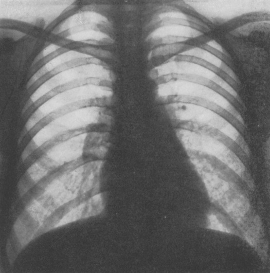
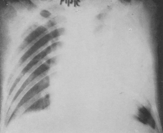
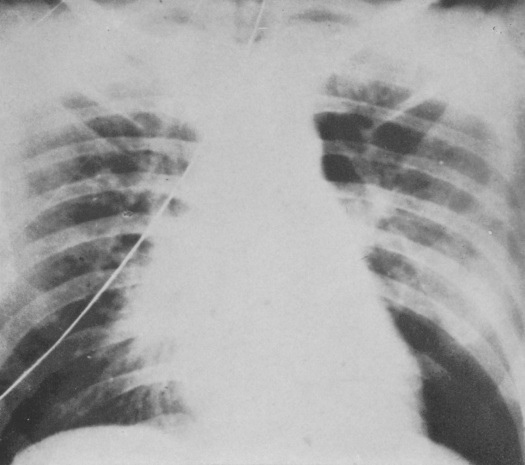
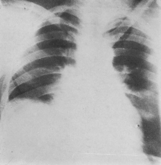
A follow-up to the Unofficial Guide to Radiology, and part of the Unofficial Guide to Medicine series, this new book The Unofficial Guide to Radiology: 100 Practice Chest X-rays, with full colour annotations and full X-ray reports  has at its heart the inspiring idea that the development of educational resources should be driven by those who use them. The result is a fantastic resource for reporting radiographers, medical students, junior doctors in any specialty, providing a comprehensive and practical approach to chest x-ray interpretation.

41Vnk61P4sL._SX352_BO1,204,203,200_
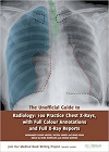
Right from the start, the book’s cover is self-explanatory and is easily perceived to be about chest X-ray interpretations.   The 100 chest X-ray cases are presented in a test-yourself format, with the images and case history presented on one page and the interpretation and report on the next.

The cases are separated in three coloured divisions: Standard (orange), Intermediate (purple) and Advanced (blue). The first page provides the reader with a short clinical indication followed by the associated chest X-ray in high quality, all in one page. The second page then evaluates the technical features, again using a colour code scheme which is then diagrammatically presented on the same chest X-ray, but on a smaller scale. It may be coincidence that the orange, purple and blue technical features can also be perceived as standard, intermediate and advanced technical points to look out for from a radiographer’s perspective. Finally, there is a short but precise summary demonstrating a report of the chest X-ray followed by further management for the patient.

The image quality is excellent in comparison to most other available textbooks, with crisp full-page images allowing the detail of the images to be explored – crucial in the days of PACS when every possible abnormality can be magnified a hundredfold.

Each ‘answer’ page has a consistent format, embedding a sensible interpretation pathway, and a clear layout highlighting both normal and abnormal findings. The consistency, and the detailed and comprehensive annotations, allows the reader to build up an idea of ‘normal’ over the course of the cases, continuously reinforcing important structures to check on every radiograph.

The multidisciplinary approach to development also comes through strongly, with suggested first management steps in response to each radiograph placing the interpretation firmly in the pragmatic clinical world. However, the ‘reporting’ style employed also develops familiarity with the language of radiologists; if this can sometimes seems overly formal or formulaic, it serves a purpose in ensuring that clinicians and radiologists are on the same page.

The clinical cases provided are realistic and are what you expect to find whether in Accident and Emergency and/or outpatient, GP clinics. From pathologies to pneumothoraxes, fractures to line insertions, most scenarios are covered in this book.

Valandis Kostas strongly recommends this book to all grade and advanced radiographers. He observes that the book provides the patient pathway link from clinical presentation to radiology, to treatment and type of follow up imaging required i.e. CT and/or chest clinic referral. The layout enables understanding of the acquired chest x-ray, vital for best practice.

He particularly applauded the section on quality of the chest X-ray, using the similar 10 point image quality check radiographers use in their clearance of X-rays they undertake. Patient I.D, rotation, penetration and inspiration are a few examples. Furthermore, the case layout educates radiographers the importance of these checks to aid image interpretation for diagnosis whilst encouraging learning about chest pathologies. This will eliminate the repetitious perception of the chest X-ray and it will encourage radiographers to maintain high quality chest radiographs for accurate diagnosis and reduce false negatives and false positives.

The clinical details provided in the case vignettes are of a level of detail that surpasses most of those seen in clinical practice; hopefully, the detail provided here will also serve to demonstrate to clinicians who read the book how fundamental these details are, and serve as a resource on helpful requesting as well as interpretation of chest radiographs.

An important area for radiographers and radiologists that is not covered in as much detail is the inadequate chest x-ray, and perhaps the book could be improved by including a few examples of misses/near misses from poor quality radiographs in order to educate readers on when a repeat X-ray is required.

Tom Campion, trainee radiologist  would happily recommend the book to anyone whose job involves X-ray reporting as it delivers a solid foundation in interpretation skills and serves  as both a thoughtfully structured introduction to the beginner and a handy reference to the more experienced.

Both Valandis and Tom felt that the book would make a great app or online tool  in the future.

The Unofficial Guide to Radiology £19.99

https://www.amazon.co.uk/Unofficial-Guide-Radiology-Practice-Annotations/dp/1910399019

Images: (Top left) Tom Campion, (top right) Valandis Kostas.

AUTHORS:

by Mohammed Rashid Akhtar MBBS BSc (Hons) FRCR (Author), Na’eem Ahmed MBBS BSc (Author), Nihad Khan MBBS BSc (Author)

EDITORS:

Mark Rodrigues MBChB(Hons) BSc(Hons) FRCR (Editor), Zeshan Qureshi BM BSc (Hons) MSc MRCPCH (Editor)


Hats off to Sir Peter Mansfield (1933-2017)

13-sir-peter-mansfield-2003

Sir Peter Mansfield left school with no qualifications to become one of the most eminent scientists in the world of physics. Here, Dr Adrian Thomas pays tribute to the man who lived through World War Two and with dogged determination forged his way in science to become a distinguished and recognised physicist who played a major part in the story of MRI.


Sir Peter Mansfield was born on 9 October 1933 in Lambeth in London, and grew up in Camberwell. His mother had worked as a waitress in a Lyons Corner House in the West End of London, and his father first worked as a labourer in the South Metropolitan Gas Company, and then as a gas fitter. Mansfield recounted being sent with other children on a holiday to Kent for disadvantaged London children by the Children’s Country Holiday Fund.

Peter Mansfield was 5 years old when the war broke out in 1939. He remembers standing with his father at the entrance of an air raid shelter watching anti-aircraft shells exploding around German bombers caught in the searchlights. As the Blitz intensified he was evacuated from the dangers of the capital, as were so many other London children. With his brother he was sent to Devon, where he was assigned to Florence and Cecil Rowland who lived in Babbacombe, Torquay. The Rowlands were called Auntie and Uncle, and Mansfield  attended the nearby junior school. Cecil Rowland was a carpenter and joiner by trade, and encouraged Peter to develop his practical skills by giving him a toolbox, and tools were slowly acquired. He obviously obtained some proficiency since with some guidance he made several wooden toys which he was able to sell at an undercover market and a toyshop in Torquay. His life was not without danger even outside London, and in early 1944,whilst out playing, he saw a German twin-engined Fokke-Wulf plane flying at rooftop level. The tail gunner was spraying bullets everywhere, and he rapidly took shelter behind a dry-stone wall.

On his return to London his secondary schooling was at Peckham Central, moving  to the William Penn School in Peckham. Shortly before he left school at 15 he had an interview with a careers adviser. Peter said that he was interested in science, and the adviser responded that since he was unqualified that he should try something less ambitious. He was interested in printing and so took up an apprentice in the Bookbinding Department of Ede and Fisher in Fenchurch Street in the City of London, and whilst there he took evening classes.   Developing an interest in rockets he was offered a position at the Rocket Propulsion Department (RPD) at Westcott, near Aylesbury.

In 1952 he was called up into the Army for his National Service, where he joined the Engineers. The Army allowed him to develop his interest in science. On demobilization he returned to Westcott and completed his A levels. This enabled him to apply for a special honors degree course in physics at Queen Mary College in London. In 1959 he obtained his BSc, and three years later he was awarded his PhD in physics. From 1962 to 1964 he was Research Associate at the Department of Physics at the University of Illinois, and in 1964 was appointed Lecturer at the Department of Physics at the University of Nottingham.

During a sabbatical in Heidelberg in 1972 Mansfield corresponded with his student, Peter Grannell in Nottingham, and became interested in what became MRI, presenting his first paper in 1973 at the First Specialized Colloque Ampère. Mansfield developed a line scanning technique, and this was used to scan the finger of one of one of his early research students, Dr Andrew Maudsley. The scan times required for these finger images varied between 15 and 23 minutes. These were the first images of a live human subject and they were presented to the Medical Research Council, which in 1976 was reviewing the work of various groups including those in Nottingham and Aberdeen.

13-terry-baines-peter-mansfield-and-andrew-maudsley-c1974

In 1977 the team at Nottingham, which included the late Brian Worthington, successfully  produced an image of a wrist. The following year Mansfield presented his first  abdominal image. In 1979 Peter Mansfield was appointed Professor of Physics at the University of Nottingham. As the Nobel Committee emphasized, the importance of the work of Peter Mansfield was that he further developed the utilization of gradients in the magnetic field. Mansfield demonstrated how the signals could be mathematically analyzed, which resulted in the development of  a practical  imaging technique. Mansfield also demonstrated how to achieve extremely fast imaging times by developing echo-planar imaging. This is all very impressive for a boy who left school at 15 with no qualifications.

13-sharing-an-amusing-tale-with-paul-lauterbur-2003

Peter Mansfield was awarded many prizes and awards including:

the Gold Medal of the Society of Magnetic Resonance in Medicine (1983); Fellow of the Royal Society (1987); the Silvanus Thompson Medal of the British Institute of Radiology (1988); the International Society of Magnetic Resonance (ISMAR) prize (jointly with Paul Lauterbur)(1992);  Knighthood (1993); Honorary Fellow of the Royal College of Radiology and Honorary Member of the British Institute of Radiology (1993);  the Gold Medal of the European Congress of Radiology and the European Association of Radiology (1995);  Honorary Fellow of the Institute of Physics (1997); the Nobel Prize for Medicine together with Paul Lauterbur (2003);   Lifetime Achievement Award presented by Prime Minister Gordon Brown (2009).

His autobiography The Long Road to Stockholm, The Story of MRI was published in 2013. This is an interesting read, particularly in relation to his early years, and is recommended reading for everyone interested in the radiological sciences. This is a revealing account of a remarkable life. Whilst we may discuss the complexities of the development of MRI and exactly who should have received the Nobel Prize, there can be no doubt about his major contributions. MRI has made, and is making major contributions to health care. He died age 83 on 8 February 2017.

The University of Nottingham has set up an online book of condolence http://www.nottingham.ac.uk/news/sir-peter-mansfield/

About Dr Adrian Thomas, Honorary Historian BIR

Dr Thomas was a medical student at University College, London. He was taught medical history by Edwin Clarke, Bill Bynum and Jonathan Miller. In the mid-1980s he was a founding member of what is now the British Society for the History of Radiology. In 1995 he organised the radiology history exhibition for the Röntgen Centenary Congress and edited his first book on radiology history.

He has published extensively on radiology history and has actively promoted radiology history throughout his career. He is currently the Chairman of the International Society for the History of Radiology.

Dr Thomas believes it is important that radiology is represented in the wider medical history community and to that end lectures on radiology history in the Diploma of the History of Medicine of the Society Apothecaries (DHMSA). He is the immediate past-president of the British Society for the History of Medicine, and the UK national representative to the International Society for the History of Medicine.

See more on the history of radiology at http://www.bshr.org.uk


Breaking the mould – how  radiographer reporting is better for the patient.

nigel-thomas

Professor Nigel Thomas from the University of Salford explains why allowing a radiographer to report X-rays  is not threat to the radiology profession.


I’ll nail my colours to the mast straight away, and state that I have been an active proponent of radiographer role extension in general, and radiographer reporting in particular, for over 20 years.

I first became involved in mid 1995 when the University of Salford (then University College Salford) asked for help in setting up a formal plain film reporting course for radiographers. The context for this was the unresolved tension between the large numbers of unreported films in most X-ray Departments and the realisation that radiographers as a group of professionals were often working below their full potential – a real untapped resource within our own departments. Becoming involved in the process seemed to me to be a very obvious thing to do, and I have never had any regrets about doing so. I don’t believe that I have contributed to the demise of my profession, and I certainly don’t feel like a “turkey voting for Christmas”.

Over the years since then, radiographers have increased the breadth of their involvement in reporting (to currently include some types of MR scanning and CT, as well as gastro-intestinal contrast studies amongst other things), as well as developing a career structure which encompasses working at Advanced Practitioner and Consultant Radiographer levels (the latter being a particular success in the world of breast imaging, where consultant radiographers can follow an entire patient journey by being able to perform and report mammograms, perform and report breast ultrasound and perform guided biopsies, as well as having counselling skills).

It was clear from the beginning that there would be opposition to the idea of radiographer reporting, both from the radiology establishment, and, to a much lesser extent, from within the radiography profession itself. In order to ensure that the process of creating reporting radiographers was as good as it could be, certain quality measures were put into place. No radiographer can report in the UK without a recognised qualification (at PgC or Pgd level) gained from a higher education institution. In the context of the workplace, reporting is done within an agreed scheme of work (signed off by the employing Trust Board), and regular audit is undertaken.

In 2017 between 15 and 20% of all plain film examinations in the UK are reported by radiographers, and there are now over 50 people in consultant radiographer grades around the country. Reporting radiographers have been “part of the furniture” in X-ray departments for over 20 years, and generations of junior doctors, nurses and physiotherapists have been familiar with using them as a port of call for advice on the interpretation of images.

And yet, despite all of the above, resistance to radiographer reporting persists. I find this particularly perplexing for several reasons:

  1. The reporting shortfall still persists, and patients are being put at risk by our failure to report their examinations in a timely and accurate way – would we rather leave them unreported?
  2. Radiologists have more than enough to do – there are too few of us, and our time is used to apply our unique skill set to report labour intensive complex examinations, undertake time-consuming interventional procedures, and provide a commitment to the support of MDTs.
  3. There is a substantial body of sound scientific evidence (published in the major UK peer-reviewed radiological journals) that radiographer reporting works, is safe, and is of a comparable standard to that provided by medical staff in many areas.
  4. Radiologists have been involved in this process from day 1 – advising on course content, giving lectures, acting as examiners and external examiners, and, most importantly, acting as mentors to radiographers in training at their places of work.

The final irony for me, as we progress into the 21st century is that, despite all the above, it is clear that some of my colleagues are much keener to gain help from computers than humans. Don’t get me wrong, I’m sure that Computer Aided Design (CAD) and Artificial Intelligence (AI)  will have a huge role to play in the routine provision of a radiology service in the near future, but reporting radiographers can help patients here and now.

References

Berman L, de Lacey G, Twomey E, Twomey B, Welch, T and Eban, R. ‘Reducing errors in the accident department: a simple method using radiographers’, British Medical Journal 1985; 290: 421-2

Loughran,C.F., Reporting of fracture radiographs by radiographers: the impact of a training programme. British Journal of Radiology, 67(802), 945 –950, 1994

Judith Kelly, Peter Hogg, Suzanne Henwood. The role of a consultant breast radiographer: A description and a reflection. Radiography, Volume 14, Supplement 1, e2-e10, 2008.

Brealey, S., Hewitt, C., Scally, A., Hahn, S., Godfrey, C., and Thomas, N.B. Bivariate meta-analysis of sensitivity and specificity of radiographers’ plain radiograph reporting in clinical practice. British Journal of Radiology, 82, (979), 600-604, 2009.

Piper, K., Buscall, K., Thomas, N.B., MRI reporting by radiographers: Findings of an accredited postgraduate programme. Radiography, Volume 16, Issue 2, 136-142, May 2010

  1. Piper, S. Cox, A. Paterson, A. Thomas, N.B. Thomas, N. Jeyagopal, N. Woznitza. Chest reporting by radiographers: Findings of an accredited postgraduate programme, Radiography, Volume 20, Issue 2, 94-99, February 2014
  1. Snaith, M. Hardy, E.F. Lewis Radiographer reporting in the UK: A longitudinal analysis

Radiography, Volume 21, Issue 2, 119-123, 2015

About Nigel Thomas

Born and raised in Cornwall, I qualified from St Bartholomew’s Hospital in London in 1981 having gained an intercalated B.Sc in Biochemistry in 1978.

My radiology training was undertaken on the North Western Training Scheme (based in Manchester), and I was appointed as Consultant Radiologist to North Manchester General Hospital in 1989.In 2005 I moved to a Consultant post at Trafford General Hospital and retired as a full-time NHS Consultant Radiologist in 2015.

I currently work as an independent Consultant Radiologist and, amongst other roles, am a mentor to Reporting Radiographers at two large Foundation Trusts in the Manchester conurbation.

I first became involved in the process of radiographer role development at the University of Salford in 1995, and was appointed as an Honorary Professor there in 2000. I have over 40 publications in scientific journals, and am a co-author of a standard textbook of Obstetric and Gynaecological Ultrasound scanning.


Image: Courtesy of Nottingham University Hospitals


Top tips for honest science messages in the media

13-kate-elliottScience is often misrepresented in the media. The BIR supports the charity Sense about Science in their call for all research to be openly and honestly reported. This year we supported one of their Voice of Young Science workshops called “Standing up for Science” held on 16 September 2016 in London.

Here, Kate Elliott, Medical Physicist at  Mount Vernon Cancer Centre was one of three lucky BIR members to attend the workshop which gave young researchers top tips and advice on how to get their scientific messages across as clearly and accurately as possible.


I hate speaking in public and even the thought of writing this article terrified me. Why then, you might ask, did I apply to go on the Standing up for Science media workshop?

I often get annoyed at the coverage of science in the media and the misuse of statistics and results. Recently, the Brexit “debate” has left me ranting at friends, and I often find myself defending junior doctors on social media. When I received the email from BIR advertising the media workshop, it struck me as an opportunity to learn what I could do to positively influence the public perception of science, and to hear first-hand from journalists about their involvement.

The first session consisted of a panel of three scientists who told us of personal experiences with the press and offered advice based on this. An example which stood out to me as a healthcare scientist was Professor Stephen Keevil’s use of the media to highlight a problem with a new EU directive on physical agents[1], which could  have caused problems for MRI. Politicians took heed of his criticism, and effected a change to the directive in Brussels. This was a great example of how the media can be used effectively to influence policy – something that is likely to become increasingly important in the next few years.

The second session was a panel of three journalists, who explained their daily process for13-standing-up-for-science-workshop-sept-2016selecting and pitching stories. Science stories are selected based on interest, accessibility, and importance. These are pitched to the editors, who decide which ones to take further. The journalists pointed out that their duty is to their audience, not to science. Unfortunately, science has to compete with news on David Beckham’s haircut. Time constraints are also a problem. They write multiple articles a day (I’m three weeks and counting on this one…), so it’s important for scientists to be available to discuss their research on the day it’s published.

The third panel was about the nuts and bolts of how to interact with the media, and recommended campaigns such as Sense about Science’s “Ask for Evidence” campaign.

I left the event with the following advice to keep in mind:

  • If you disagree with something: speak out. If the public only hears one side of the story, that’s the side they’ll believe.
  • Stick to a few key points. Get those across, even if it means having to ignore questions or turn them around in an infuriatingly politician-like way!
  • Be available. If you’ve put out a press release, you need to be able to respond quickly. Journalists work to very stringent time scales, so being available in a week’s time is going to be too late.
  • Talk to the public. Attend events such as Pint of Science, or become a STEM ambassador, because that will really help you learn to speak in layman’s terms and get you used to answering obscure questions.
  • Get training. If not full media training, a workshop like this is a really good way to be slightly more prepared – and you get to hear about all the interesting science other people are involved in!

Image: BIR members  Jim Zhong, Kate Elliott and Maureen Obioha Agwanihu who attended the workshop

[1] https://www.myesr.org/html/img/pool/MRI-Report-Stephen-Keevil.pdf