

Stewart Whitley reflects on how technology has revolutionised radiographic imaging.
Since the launch of RAD Magazine back in 1975, radiographic imaging as we know it has changed dramatically, far beyond the concept of what anyone could have imagined at that time. And just as smart mobile phone technology has revolutionised how we communicate, so too has the emergence of digital imaging technology transformed the X-ray department while at the same time providing both regional and national connectivity.

Figure 1: At work in the chest room at New Ealing Hospital, London. From RAD Magazine, July 1979
A few of us will remember with fondness those ‘bygone days’ when the darkroom was a hive of activity and was central to all that happened in the X-ray department; all permanent images, and for that matter, reporting was dependent on film/screen technology and film processing chemistry. Back then there was the gradual but necessary progression from manual processing, with those famous drying cabinets, to the first automatic dryers and then the emergence of automatic processing which was the first step in revolutionising film processing and the eventual demise of the darkroom. Even though those wonderful automatic film processors could eventually process film in 90 seconds, a great deal of care and attention was still necessary to keep rollers, processing tanks and processing chemicals in tip-top condition. And what department was without a silver recovery system to generate income? Then everything changed dramatically overnight with the introduction of daylight processing. Different manufacturers had different solutions but the overall effect was to transform the X-ray department and free up the darkroom technician, many of whom became X-ray helpers – the forerunners to the modern image support worker (figure 1). While image acquisition using modern film/screen technology progressed steadily with the introduction of more efficient and higher quality image systems, the focus was on radiation dose reduction, with X-ray manufacturers offering a range of general X-ray and fluoroscopic systems which provided welcome features to reduce patient and staff dose.

Figure 2: Radiologists and radiographers attending a preview of Agfa Gevaert’s daylight processing system in London. From RAD Magazine, March 1977
Older X-ray systems were powered with what would be considered today outdated X-ray generator technology and X-ray tube design, with corresponding limitations on short exposure times and geometric sharpness. Thanks howeverto consistent research and development in generator technology and X-ray tube design, the problem of high tube output and short exposure times with associated production of inherent high heat was resolved. This facilitated multiple exposure equipment for cardiovascular imaging and general angiography with their inherent demands for high quality sharp images at low radiation doses. Such changes have enabled the acquisition of motion-free images of the vascular tree, coronary vessels and heart anatomy, giving spectacular images of cardiac function and anatomy. The X-ray generator control desk is now hardly recognisable from those found in departments back in 1975 – some still had voltage compensation controls and meters for you to manipulate before you started the day (figure 2).
Gone are those massive exposure control dials for individual control of Kv, Ma and time. Such control desks were large and floor standing, unlike modern small desks which rest on a bench or can be wall mounted and synchronised to the X-ray tube housing/light beam display unit. For exposure factor selection, we are no longer confined to manual selection, thanks to the development of anatomical programming selection combined with the introduction of automatic exposure control – something that we take for granted nowadays – but its use still requires skill and knowledge of the location and use of the relevant ionizing chambers to select the most appropriate exposure conditions. Used correctly, image quality will be consistent with the optimum use of radiation dose. The design of X-ray tables and ceiling tube suspension systems has been a gradual process, developing from simple solutions to fully integrated motorised units where preprogramming of the location of the X-ray tube/table of a vertical Bucky is linked to the body part selected for examination, requiring less effort from the radiographer in positioning heavy equipment.
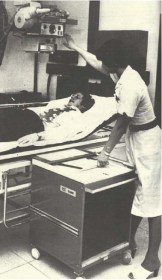
Figure 3: Coventry and Warwickshire Hospital’s ceiling-mounted equipment in its new X-ray unit. From RAD Magazine
We now see the control of exposure factor selection built in to the modern X-ray tube housing/light beam diaphragm display unit. This saves a great deal of time and releases more time for patient care, which has been further enhanced with the introduction of rise and fall tables with floating table tops – something which is taken for granted compared to the old days with fixed-height tables and no facility to move the patient other than brute force (figure 3). Overall, the advances in design with improved ergonomics have been complemented with a range of dose information and dose saving features such as the introduction of DAP meters (now a feature of all X-ray systems), additional selectable X-ray tube filtration for paediatric radiography, and the ability to remove grids in the Bucky systems to lower patient dose.
Over the years, changes in standard radiography requests and techniques have emerged which have been driven by the introduction of new technologies and patient pathways. No longer, for instance, are those well-loved isocentric skull units required because basic skull radiography has become a thing of the past and, if necessary, is replaced with the use of CT. As a result, there has been a loss of this skill, but as one modality is lost others like OPG and cone-beam computed tomography (CBCT) have found their way into the X-ray department. Continuing this theme, fluoroscopy procedures such as barium enema and barium meal procedures are no longer in favour, compared to yesteryear when they were undertaken mostly on equipment based on the undercouch X-ray tube design with over-the-table image intensifier. Not only have such fluoroscopy units in the UK diminished in number but they have been replaced with equipment with a more X-ray tube and image detector unit. This is complemented by a range of image selection features such as digital subtraction and road mapping for angiography, as well as a number of exposure and dose control options from the main control console or on a mobile control desk that can be positioned anywhere in the room.

Figure 4: Blackpool Victoria Hospital’s Farage Unit equipped with a new Philips C-arm angiography unit with CBCT capability
Such C-arm systems can also support CBCT. This truly is a leap forward in design and capability, with such configurations providing volumetric CT capabilities which in the angiography suite provide the clinician with a 3D orientation of pathology as well as a feature to plan the optimum orientation for positioning a biopsy needle, without damaging vital organs or arteries (figure 4). Undoubtedly, however, the introduction of digital technology has transformed how we acquire images. The development of both computed radiography (CR) and direct digital radiography (DDR) has been fascinating to observe. In the early days of this development, DDR with large detectors was mostly fixed and integrated into the vertical Bucky and table design while CR was based mainly on conventional cassettes, thus giving the radiographer greater flexibility and the ability to undertake examinations in the conventional way. However, all of that has changed with DDR now presented with mobile flat detectors, built-in wi-fi technology, and in different sizes capable of being used in a similar way to film/screen cassette radiography. This has revolutionised the speed in which images are acquired and, with the development of mobile DDR based X-ray systems, its use in high dependency patient care units such as ITU and SCBU is providing the clinician with instant images, thus assisting them to make immediate and important treatment decisions. Overall the X-ray department has been changed forever – what next?
This article was first published in RAD Magazine, 43, 500, 22, 24. Reproduced with permission.
About Stewart Whitley

Stewart undertook his radiography training in the Royal Army Medical Corps qualifying in 1967 at the Royal Herbert Hospital, Woolwich, London. After serving in the Army he returned to N. Ireland working first at the Lagan Valley Hospital, Lisburn and then at the Royal Victoria Hospital, Belfast where he qualified as a Radiographer Teacher before moving to Altnagelvin Hospital, Londonderry as Deputy Superintendent Radiographer.
In 1978 he was appointed District Radiographer at Blackpool Victoria Hospital where he remained until the autumn of 2006 when he retired from the NHS as Directorate Manager of Radiology and Physiotherapy Services.
Shortly after leaving the NHS he established UK Radiology Advisory Services, a small company dedicated to providing medical imaging advice and support to various NHS and private sector organisations and educational establishments.
Stewart has a passion for Radiography and his professional body, the Society and College of Radiographers, and has served as a Council Member, Honorary Secretary of the N. Ireland Branch of the Society of Radiographers and as a DCR and HDCR Medical Photography examiner as well as serving on a number of SCOR committees.
He lectures on a number of courses and was an Honorary Lecturer and Coordinator for radiographer lecturers on the FRCR course at Manchester University.
Stewart took on the role of ISRRT’s Director of Professional Practice in April 2018

As you mentioned, there are many tech advances that have helped speed up the time in which doctors can get x-ray results. One of those advances you talk about is getting the pictures digitally. I bet this has cut out much of the problems of film, and having to have a dark room. It has been awhile since I have had an x-ray, it would be interesting to see if I would notice any changes.