When medical physicists wore white coats

NHS

Dr Edwin Aird shares his memories of the revolution he has experienced working in the medical physics department.


Edwin Aird portrait

While I was an undergraduate at Newcastle University (1962), the 2nd year honours group was invited to visit the Medical Physics Department at Newcastle General Hospital. I was so impressed with the department and the range of things they were doing and the application of physics to medicine that I wrote to Professor Frank Farmer (Head of Department at that time) to ask about working there. He responded with a proposal that I apply for a special research grant that he was hoping for locally and that I write again after my graduation.

The following year I got in touch with Frank Farmer again and was accepted on an MSc grant to study “In-homogeneities in Radiotherapy”; one year in the first instance (on a grant of about £700).

Meeting 1970 1

I can’t now remember my first day exactly; not sure what office I had, but I do remember the Professor’s insistence on donning a white coat (provided and laundered by the hospital) when arriving at work, This ‘uniform’ was thought to be a vital sign to patients that physicists were part of the ‘clinical’ team.

I divided my time in that first year between research and clinical work (interestingly, this was part of the philosophy of some of the early physicists, many of whom in the 1930s and 40s had transferred from academic physics, including Frank Farmer); the situation now couldn’t be more different.

Fig 1

Image 1 The gantry mounted linear accelerator at Newcastle General Hospital [ref 2]

So, on the routine side, I began to learn about radiotherapy: its planning and treatment. Newcastle at that time had two cobalt machines (Mobaltron-60s) and two linear accelerators as well as two Marconi 250kV sets and a Philips SXT (Superficial X-rays). This latter set I used extensively a little later in my career to optimise the characteristics of the Farmer chamber (image 1).

The main linear accelerator was manufactured by Mullard (a branch of Philips), which Frank Farmer had installed as the first gantry-mounted 4MV linac (image 2). There were a few stories about the installation of this linac. In particular I remember this: the team had reached the point where they needed to produce X-rays so needed a high atomic number transmission target. Someone found a sovereign, but forgot the amount of heat produced when the high energy electrons hit it. The sovereign promptly melted and fell out of the linac head. (This linac went on to perform 25 years’ service).

Fig 2

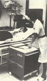
Fig 2 Newcastle Simulator (Frank Farmer at Controls) [ref 3]

For those physicists who find quality assurance (QA) on linear accelerators today a huge burden, my recollection of the checks we performed on this linac amounted to: dose measurements and field size checks. There was little attempt to measure flatness since the accelerated electron beam wasn’t bent before striking the target, so no perturbations in intensity across the field were expected.


Fig 3

Fig 3 A Head and Neck Xeroradiograph showing potential. Treatment volume [ref 3]


Treatment planning was done by hand, often mainly using % depth dose, but also with some isodose curves for more complex plans on tracing paper; on to an outline of the patient performed using lead wire and other devices, e.g. callipers. Newcastle was unique in having developed a home-made simulator (using a radiotherapy SXT unit mounted on a gantry (image 2) that allowed an image to be viewed on a Xerox plate immediately without the need for film development (ref 3, see also image 3).

The first computer in the department, which was to revolutionise treatment planning, came to Newcastle in the early 1970s and was called a PDP 8, the ‘Rad8 system’ developed by Bentley and Milan. (image 4 [ref 4]).

Radium tubes and needles were still extensively used at this time for intracavitary and interstitial brachytherapy. I was also required to calculate the dwell times for gynae radium insertions (using ovoids and central vaginal sources with a modified Manchester system for the gynae and radium needles, mainly in back of tongue with radiographs and Paterson Parker tables). Following the calculation of the radium dwell times I would then go up to the ward to discuss the removal times, for each patient, with sister on the ward. Amazing now to think how much radioactivity (up to 105mg – equivalent to approximately 389 MBq-or more) for gynae insertions was handled by several different groups of staff at that time.

In those early years I also learnt the elements of radiation protection and nuclear medicine. Radiation Protection (guided by a ‘Code of Practice’; an excellent document that was used to help write the ‘Guidance Note’ now used under Ionising radiation Legislation) was regionally organised. Very early in my career (there were only two of us to perform the external radiation work: myself and MJ Day), I found myself organising visits within Northumberland, Cumbria and parts of Durham. (See The Regional Centre below). These were more as inspections than to make many measurements (not the very extensive QA that is performed now, although we did use film or image intensification – only recently developed – to look for faults in lead aprons; and we measured exposure levels in and out of beam with the ‘37D’ (Pitman) dosemeter (originally developed by Sidney Osborne as an excellent versatile instrument for exposure measurements ref 5). I learnt a pattern of inspection of barriers, filtration, lead aprons etc. very quickly. [In the early 1970s the NRPB decided to do their own inspections and were surprised to find that, because the HPA had organised things so well there was little need for NRPB to get too heavily involved].

My memories of nuclear medicine: incredibly slow rectilinear scanners, prior to the commercial development of gamma cameras; kidney function (renogram) using two ‘D’-shaped detectors (scintillation) with manual optimisation of their positions connected to count rate meters and chart recorders.

The regional centre

I’m not sure how many hospitals this covered in my early days in Northumberland, Tyneside, Durham and Cumbria. I know when Keith Boddy implemented Frank Farmer’s plan to have a small physics department (where gamma cameras were installed in district general hospitals) there were thirteen hospitals with physics departments in the region. Prior to this – from about my 3rd year at Newcastle – I was delegated to look after the small centre in Carlisle where there were two Marconi 250kV sets, an SXT and some iodine treatment. In the 1950s this had been the job of Jack Fowler who used some of his time to study arc therapy on 250kV (see ref 6).

Rad 2014 photo

Edwin Aird (right) receiving the BIR Sylvanus Thompson Award from Andy Beavis

In those early years I also found time to help develop differential X-ray absorptiometry to measure antimony in the local Tyneside Antimony workers’ lungs (organised by the Newcastle University Department of Industrial Health). I was also able to build on this experience to develop my own equipment, using characteristic X-rays, to measure bone mineral in the femur. This clinical work allowed me to meet a new set of clinicians (other than radiotherapists – as Clinical Oncologists were then called – and radiologists) involved with bone loss in patients: kidney specialists, endocrinologists and geriatricians. Commercial equipment has since been developed to perform bone mineral and body composition measurements (GE Lunar, Hologic, and Norland).


References:

  1. Aird EGA and Farmer FT. The design of a thimble chamber for the Farmer dosemeter. Phys Med Biol 1972 17: 169–174. https://doi.org/10.1088/0031-9155/17/2/001
  2. Day MJ and Farmer FT. The 4 MeV Linear Accelerator at Newcastle upon Tyne. Br J Radiol 1958; 31: 669–682. https://doi.org/10.1259/0007-1285-31-372-669
  3. Farmer FT, Fowler JF and Haggith JW. Megavoltage Treatment Planning and the Use of Xeroradiography. Br J Radiol 1963; 36: 426–435. https://doi.org/10.1259/0007-1285-36-426-426
  4. Bentley RE and Milan J. An interactive digital computer system for radiotherapy treatment planning. Br J Radiol 1971; 44: 826–833. https://doi.org/10.1259/0007-1285-44-527-826
  5. Osborn SB and Borrows RG. An Ionization Chamber for Diagnostic X-Radiation. Med.Biol1958; 3: 37–43. https://doi.org/10.1088/0031-9155/3/1/305
  6. Fowler JF and Farmer FT. Measured Dose Distributions in Arc and Rotation Therapy: A Critical Comparison of Moving and Fixed Field Techniques. Br J Radiol 1957; 30: 653–659. https://doi.org/10.1259/0007-1285-30-360-653

About Dr Edwin Aird

Edwin Aird was Head of Physics at Mount Vernon Hospital from 1988-2012 and Head of Radiotherapy Physics at St Bartholomew’s Hospital from 1985-1988. He was Head of Radiotherapy Physics at Newcastle General Hospital from 1980-1985. He is an IPEM Chief Examiner 1994-1997 (Physics Training Scheme), FRCR (RT) Examiner 1989-1993, Radiotherapy Degree (External Examiner (Liverpool University): 1993-1996. He is a IAEA: Qualified Expert: 1999-2005 and an LH Gray Trustee: 1999-2003.

He was awarded the BIR Roentgen Prize in 2005 and delivered the Silvanus Thompson: Award and Eponymous Lecture 2013.

Having a scan with your head in a rubber hat

NHS

Dr Jim Stevenson, reflects on life as a radiologist in the 1970s.


Jim StevensonI started my radiological life in the mid seventies at St George’s Hospital. Part of the rotation programme involved some time at the Atkinson Morley Hospital where I came across the first generation scanner. There was an old dental chair on which a patient laid back with his head in a rubber hat in the scanner porthole. It took 8 slices. Each slice took 5 minutes using an old fashioned tomogram X-ray tube. The image details were processed by a very large computer. The resultant image was printed on a photograph. The image matrix was 80 by 80, an advance since the original 40 by 40. How Jamie Ambrose invented the reports I do not know but his detailed knowledge of brain anatomy was quite outstanding.

Once when walking past the scanner I saw a porter in a brown overall walking round the machine. Being concerned about security, I spoke to Jamie Ambrose. “Don’t worry about him,” he said, ‘”That’s only Godfrey“ (Hounsfield from EMI).

Significant advances in CT occurred about every 5 years. When the first body images appeared we all had to learn cross-sectional anatomy. Since 1945 all anatomy was taught in longitudinal section – sagittal and coronal. I showed an image to my father-in-law. He had no problem with it but he had qualified in 1940. Before the war, all medics had to learn cross-section anatomy! The very best cross-section anatomy book I found was Eycleshymer and Schoemaker published in America in 1911. Still much better than the modern ones of recent times. The only difficulty is that all the labels are in Latin which can make interpretation difficult!

Over the past fifty years medical technology advances have been and will continue to be outstanding. The need to make proper use of them hasn’t changed. Wet films, fluorescent imaging, U/S, MRI and digital are all contributing to our future.


About Dr Jim Stevenson

Dr James Duncan Stevenson BSc. MB.BS, FRCR trained at St.Thomas’ Hospital Medical School, London and four years later turned to radiology at St.George’s Hospital, London. In November 1980 he became a Consultant Radiologist at Royal Victoria Hospital, Bournemouth and Poole Hospital. He retired in August 2007.

From darkroom to digital: Tracing the transformation of Radiography

NHS

Stewart Whitley reflects on how technology has revolutionised radiographic imaging. 


Since the launch of RAD Magazine back in 1975, radiographic imaging as we know it has changed dramatically, far beyond the concept of what anyone could have imagined at that time. And just as smart mobile phone technology has revolutionised how we communicate, so too has the emergence of digital imaging technology transformed the X-ray department while at the same time providing both regional and national connectivity.

Fig 1

Figure 1: At work in the chest room at New Ealing Hospital, London. From RAD Magazine, July 1979

A few of us will remember with fondness those ‘bygone days’ when the darkroom was a hive of activity and was central to all that happened in the X-ray department; all permanent images, and for that matter, reporting was dependent on film/screen technology and film processing chemistry. Back then there was the gradual but necessary progression from manual processing, with those famous drying cabinets, to the first automatic dryers and then the emergence of automatic processing which was the first step in revolutionising film processing and the eventual demise of the darkroom. Even though those wonderful automatic film processors could eventually process film in 90 seconds, a great deal of care and attention was still necessary to keep rollers, processing tanks and processing chemicals in tip-top condition. And what department was without a silver recovery system to generate income? Then everything changed dramatically overnight with the introduction of daylight processing. Different manufacturers had different solutions but the overall effect was to transform the X-ray department and free up the darkroom technician, many of whom became X-ray helpers – the forerunners to the modern image support worker (figure 1). While image acquisition using modern film/screen technology progressed steadily with the introduction of more efficient and higher quality image systems, the focus was on radiation dose reduction, with X-ray manufacturers offering a range of general X-ray and fluoroscopic systems which provided welcome features to reduce patient and staff dose.

Fig 2

Figure 2: Radiologists and radiographers attending a preview of Agfa Gevaert’s daylight processing system in London. From RAD Magazine, March 1977

Older X-ray systems were powered with what would be considered today outdated X-ray generator technology and X-ray tube design, with corresponding limitations on short exposure times and geometric sharpness. Thanks howeverto consistent research and development in generator technology and X-ray tube design, the problem of high tube output and short exposure times with associated production of inherent high heat was resolved. This facilitated multiple exposure equipment for cardiovascular imaging and general angiography with their inherent demands for high quality sharp images at low radiation doses. Such changes have enabled the acquisition of motion-free images of the vascular tree, coronary vessels and heart anatomy, giving spectacular images of cardiac function and anatomy. The X-ray generator control desk is now hardly recognisable from those found in departments back in 1975 – some still had voltage compensation controls and meters for you to manipulate before you started the day (figure 2).

Gone are those massive exposure control dials for individual control of Kv, Ma and time. Such control desks were large and floor standing, unlike modern small desks which rest on a bench or can be wall mounted and synchronised to the X-ray tube housing/light beam display unit. For exposure factor selection, we are no longer confined to manual selection, thanks to the development of anatomical programming selection combined with the introduction of automatic exposure control – something that we take for granted nowadays – but its use still requires skill and knowledge of the location and use of the relevant ionizing chambers to select the most appropriate exposure conditions. Used correctly, image quality will be consistent with the optimum use of radiation dose. The design of X-ray tables and ceiling tube suspension systems has been a gradual process, developing from simple solutions to fully integrated motorised units where preprogramming of the location of the X-ray tube/table of a vertical Bucky is linked to the body part selected for examination, requiring less effort from the radiographer in positioning heavy equipment.

Fig 3

Figure 3: Coventry and Warwickshire Hospital’s ceiling-mounted equipment in its new X-ray unit. From RAD Magazine

We now see the control of exposure factor selection built in to the modern X-ray tube housing/light beam diaphragm display unit. This saves a great deal of time and releases more time for patient care, which has been further enhanced with the introduction of rise and fall tables with floating table tops – something which is taken for granted compared to the old days with fixed-height tables and no facility to move the patient other than brute force (figure 3). Overall, the advances in design with improved ergonomics have been complemented with a range of dose information and dose saving features such as the introduction of DAP meters (now a feature of all X-ray systems), additional selectable X-ray tube filtration for paediatric radiography, and the ability to remove grids in the Bucky systems to lower patient dose.

Over the years, changes in standard radiography requests and techniques have emerged which have been driven by the introduction of new technologies and patient pathways. No longer, for instance, are those well-loved isocentric skull units required because basic skull radiography has become a thing of the past and, if necessary, is replaced with the use of CT. As a result, there has been a loss of this skill, but as one modality is lost others like OPG and cone-beam computed tomography (CBCT) have found their way into the X-ray department. Continuing this theme, fluoroscopy procedures such as barium enema and barium meal procedures are no longer in favour, compared to yesteryear when they were undertaken mostly on equipment based on the undercouch X-ray tube design with over-the-table image intensifier. Not only have such fluoroscopy units in the UK diminished in number but they have been replaced with equipment with a more X-ray tube and image detector unit. This is complemented by a range of image selection features such as digital subtraction and road mapping for angiography, as well as a number of exposure and dose control options from the main control console or on a mobile control desk that can be positioned anywhere in the room.

Image 4

Figure 4: Blackpool Victoria Hospital’s Farage Unit equipped with a new Philips C-arm angiography unit with CBCT capability

Such C-arm systems can also support CBCT. This truly is a leap forward in design and capability, with such configurations providing volumetric CT capabilities which in the angiography suite provide the clinician with a 3D orientation of pathology as well as a feature to plan the optimum orientation for positioning a biopsy needle, without damaging vital organs or arteries (figure 4). Undoubtedly, however, the introduction of digital technology has transformed how we acquire images. The development of both computed radiography (CR) and direct digital radiography (DDR) has been fascinating to observe. In the early days of this development, DDR with large detectors was mostly fixed and integrated into the vertical Bucky and table design while CR was based mainly on conventional cassettes, thus giving the radiographer greater flexibility and the ability to undertake examinations in the conventional way. However, all of that has changed with DDR now presented with mobile flat detectors, built-in wi-fi technology, and in different sizes capable of being used in a similar way to film/screen cassette radiography. This has revolutionised the speed in which images are acquired and, with the development of mobile DDR based X-ray systems, its use in high dependency patient care units such as ITU and SCBU is providing the clinician with instant images, thus assisting them to make immediate and important treatment decisions. Overall the X-ray department has been changed forever – what next?

This article was first published in RAD Magazine, 43, 500, 22, 24. Reproduced with permission.


About Stewart Whitley

Stewart Whitley

Stewart undertook his radiography training in the Royal Army Medical Corps qualifying in 1967 at the Royal Herbert Hospital, Woolwich, London.  After serving in the Army he returned to N. Ireland working first at the Lagan Valley Hospital, Lisburn and then at the Royal Victoria Hospital, Belfast where he qualified as a Radiographer Teacher before moving to Altnagelvin Hospital, Londonderry as Deputy Superintendent Radiographer.

In 1978 he was appointed District Radiographer at Blackpool Victoria Hospital where he remained until the autumn of 2006 when he retired from the NHS as Directorate Manager of Radiology and Physiotherapy Services.

Shortly after leaving the NHS he established UK Radiology Advisory Services, a small company dedicated to providing medical imaging advice and support to various NHS and private sector organisations and educational establishments.

Stewart has a passion for Radiography and his professional body, the Society and College of Radiographers, and has served as a Council Member, Honorary Secretary of the N. Ireland Branch of the Society of Radiographers and as a DCR and HDCR Medical Photography examiner as well as serving on a number of SCOR committees.

He lectures on a number of courses and was an Honorary Lecturer and Coordinator for radiographer lecturers on the FRCR course at Manchester University.

Stewart took on the role of ISRRT’s Director of Professional Practice in April 2018


The case of the missing fingers!

NHS

Professor Roger Dale remembers how he got his first job in medical physics and how he thought he’d discovered a radiation martyr.


Roger Dale circa 1966

Anxiously seeking a job in medical physics on completion of my first degree in 1966 I quickly became aware that basic grade physicist positions in large centres were difficult to find and, for a while, I was unsure what to do. Being out of work I wrote in some desperation to a (very small) radiotherapy centre in Kent pointing out my predicament and asking if I could join as a porter until such time as I could obtain a physicist position in a larger department. To my great surprise I received a phone call a day or two later from the head radiotherapist (Dr B) inviting me along for an informal chat with him, during which it transpired that the hospital had no requirement for any more porters but did have a vacant establishment for a radiotherapy physicist at principal grade! The principal post had already been offered to a gentleman in New Zealand but it would take a month or two before he could take up the position. Therefore, as there was no physicist in post at that time, Dr B suggested that I join as an acting-temporary(!) basic grade until the principal appointee arrived in the UK. Needless to say, I agreed without hesitation.

The necessary paperwork was sorted out remarkably quickly (the old personnel departments always seemed notably more efficient than the burgeoning HR empires which later followed) and my career in medical physics began, albeit rather shakily. My only ‘supervision’ came from occasional conversations with Mr W, the Chief Technician, whose own duties were entirely focused on running the film badge and thyroid uptake services. He was not at all involved on the radiotherapy side of things so I spent many hours buried deep in the standard radiotherapy physics textbooks of the time. That reading reinforced my desire to stay in medical physics because here were the seemingly abstract physical and mathematical concepts encountered during my degree studies being successfully applied to highly relevant clinical issues. Amongst other things I brushed up on the fundamentals of radium dosimetry, this being necessary since Dr B performed several radium implants each week (remote afterloading systems were only just being introduced back then) and, as I was now the sole medical physicist (of sorts) within a 50 mile radius, he required me to be present during the procedures.

Dr B’s theatre sessions were an eye-opener. Apart from a certain squeamishness at witnessing surgery for the first time, I found his implantation technique quite scary since, although a full range of surgical implements and manipulators were at his disposal, he had a habit of giving all the radium needles a push with his fingers. Worse, it was impossible not to notice that several of his fingers were in fact missing! Even a greenhorn like me knew that physically touching radioactive sources was definitely a practice not to be recommended and the fledgling scientist in me began to ponder on cause and effect.

For several days it worried me that Dr B might be paying a very high price in order to pursue his noble vocation and I was unsure how (or if) I should air my concerns, especially as my status as an unsupervised acting-temporary basic grade physicist of just a few weeks’ standing hardly conferred much authority. Eventually I plucked up the courage to speak to the Chief Technician, telling him how convinced I was that Dr B was suffering radiation damage as a direct result of his operating technique. Mr W’s reaction was not quite what I expected. After some snorts of derision at my expense he then took some delight in pointing out that Dr B had been in the RAMC during the war. He had landed on the Normandy beaches where his jeep had hit a mine, and that was how he had lost several of his fingers. Somewhat chastened, I went away to reflect on the fact that my powers of deductive reasoning might be in need of substantial refinement.

Shortly after this awkward conversation the newly-appointed principal physicist arrived from New Zealand and, contrary to all my expectations, Dr B suggested that I stay on for a while longer to gain some first-hand experience working with the new man. This was to be a tremendous bonus as the knowledge and advice I picked up in the weeks following gave me enough of an advantage to successfully apply for a substantive post (i.e. neither acting nor temporary) in a large London centre, after which I never looked back.

Roger Dale recentToday’s NHS is nothing like the one I joined in 1966 and specialised scientist training is much more formalised and incalculably better. No one these days could be appointed in the manner that I had been but Dr B, like most other NHS professionals then and now, was motivated by good intentions and his thoughtfulness over fifty years ago put me on the path to a rich and fulfilling career in medical physics and radiobiology. I discovered later in life that Dr B had told one of his colleagues that he had helped me because he “wanted to give the lad a chance”. What he gave me was a chance that was truly exceptional and this lad has been immensely grateful ever since.


About Professor Roger Dale

Roger Dale retired from his post at Imperial College Healthcare in 2010 following an NHS career spanning 43 years. His main scientific interest has been the development of radiobiological models which can be used to quantitatively assess the biological impact of radiotherapy and other cancer treatment modalities. He is widely published and the clinical significance of his work has been recognised through the award of a number of prestigious scientific prizes and through his  parallel appointment, in 2005, as Professor of Cancer Radiobiology in the Faculty of Medicine at Imperial College. He continues to be involved in research and teaching.

My first day in radiotherapy physics: reflections of a medical physicist

NHSIn 2010 Karen Goldstone was awarded the MBE for her services to healthcare. Here she reflects on the primitive tools used for radiotherapy patient outlines back in the 1970s and remembers the wise advice she was given on her first day as a radiotherapy physicist.

BIR

I started work in the NHS as a Hospital Physicist in 1970. Prior to that I did the MSc in radiation Physics based at Middlesex Hospital. When doing a placement in nuclear medicine, computer tapes had to be taken to University College about a fifteen minute walk to the other side of Tottenham Court Road and fetched the next day hopefully having run successfully.

In my first post I expected to be doing mainly diagnostic radiology physics but discovered that that was rather a luxury field and so most of my time was spent doing radiotherapy physics. Those were the days when patient outlines were taken using a strip of lead or a flexicurve and planning was done using tracing paper and coloured pencils or biros. There was no computer planning of course and we only had one calculator with a paper roll print out so slide rules were in constant use. The main piece of advice I remember receiving on my first day was that if I discovered I had made a mistake I should own up to it straight away and not seek to cover it up – very wise words.

When not doing radiotherapy physics many hours were spent reading out film densities produced using our homemade “Ardran Cassette” in order to check kVp. This was the beginning of setting up a quality control programme for X-ray units. Another time-consuming activity was sealing lithium borate powder into plastic capsules in order to measure dose to radiologists, carrying out various procedures under fluoroscopic control, and subsequently reading the doses received.

Although diagnostic radiology physics was not seen as important it was an exciting time and I was fortunate enough to hear Godfrey Hounsfield give the 1972 MacRobert Award lecture on “Computerised Transverse Axial Tomography” – an invention that has revolutionised diagnostic imaging.

I started my second post in 1974 in a smaller department but with responsibilities in other, far-flung, hospitals. Here I was the radiotherapy physicist (the only one) and also covered diagnostic radiology and radiation protection, but because it was a smaller department and staff had to be versatile I also did some nuclear medicine and even once some ultrasound.

In the peripheral hospitals in my patch one was still using wet developing, one using just a fluorescent screen for fluoroscopic procedures and one an image intensifier viewed not via a camera but via a mirror arrangement.

How times have changed!


About Karen Goldstone MBE

I worked for forty years in the NHS, in radiotherapy physics, diagnostic X-ray physics and all aspects of radiation protection. In 1983 I set up the East Anglian Regional Radiation Protection Service (EARRPS) based at Addenbrooke’s Hospital in Cambridge, and ran it for almost 30 years. I was both a Radiation Protection Adviser and Laser Protection Adviser. I gave physics lectures to radiologists and was a physics examiner for FRCR both in the UK and Malaysia. With colleagues in EARRPS and Cambridge University I ran a number of Radiation Protection Supervisor courses and gave countless IRMER courses to reluctant clinical staff. I was exceedingly surprised to be awarded the MBE in 2010 for services to healthcare.

Since retiring I have taken up rowing and become a Level 2 rowing coach; I have given two courses on Radiation and Health to the University of the Third Age in Cambridge and am otherwise kept busy with my garden, allotment, grandchildren and church activities. I am still involved on one or two committees for medical physics and radiological protection.

My first radiology job in the NHS

NHS

What does a jazz band, a ghost train and a figure in dark goggles have in common? They are all part of the NHS 70 memories of Professor Ralph McCready.

Ralph McCready

As a houseman I had the privilege of working for Professor Frank Pantridge, inventor of the defibrillator. I was fascinated by his catheter lab with the combination of physiology and radiology. So I decided to become a radiologist but was advised to go to England (from Northern Ireland) and obtain an impressive degree so that I could return if I wished. So I went to Guy’s Hospital, London to study for an MSc in Radiation Physics and Biology and the Diploma in Medical Radiodiagnosis (DMRD), paying my own fees.

Guy’s Radiology Department was interesting. The radiology chief was Dr Tom Hills who smoked cigars, had a tiny lead apron over the appropriate parts and had made an automatic wet X-ray film processing system.

It was obvious I would never get a radiology job at Guy’s coming from Belfast, speaking strangely, and not having the MRCP (Membership of the Royal College Physicians examination) so I applied for a Senior House Officer (SHO) position at the Hammersmith Hospital London where everybody was equal.

At the Hammersmith I was told by the other applicants that I would not get the job as I had come from Belfast. However I was determined to leave the interview with my head held high. I was first in to the SHO interview and was amazed to see a long row of people on the other side of the table headed by Professor Robert Steiner. He opened the questioning by asking why I was a member of the Musician’s Union. I explained that all my colleagues in the White Eagles Jazz Band had failed their exams, left the University and turned professional. To continue to play with them I had to join the Union. Then I was asked what else I had done, so rising to the occasion I told them I had been the ghost in a ghost train in an Amusement Park. I was bored so I connected the light over the skeleton to be permanently on. The little children came out saying that there was a ghost reading the Daily Telegraph beside the skeleton. Of course nobody believed them and the people outside poured in to see what was going on.

I emerged from the interview after forty minutes to tell the other candidates how awful the interview had been. I was appointed to the position! Professor Steiner used me to do all the odd jobs in the X-ray department for the next two years. As the junior doctor I worked in the dark with the oldest Watson X-ray set. Every time I took an erect X-ray the large steel edged cassette containing the film would slide across and usually fall out of the carriage landing on the floor with a loud crash frightening everybody in the darkened room.

It was a time of great innovation at the Hammersmith: the first renal transplant was carried out; micturating cystograms were started. After initial problems with old ladies standing up in the dark being unable to ‘pee’ when the urine hit the steel bucket with a tinkle, the problem was solved by lining the bucket with sound deadening polythene. Friday was ladies’ day when I was the only radiologist who performed Hysterosalpingography. It was done in a small room with a boiling water sterilizer in the corner. When I came out to view the films the steam poured out of the door and I would appear in a cloud of steam as a fearsome figure wearing large dark goggles and a long lead apron to the consternation of the waiting mixture of NHS and private practice ladies.

Professor Steiner was a great leader and inspiration. I will always be grateful to him appointing me to a job in the Hammersmith to start my career in the NHS. https://www.rcr.ac.uk/college/obituaries/professor-robert-steiner


About Ralph McCready

I graduated in Medicine from Queen’s University Belfast and then worked as a Houseman in the Royal Victoria Hospital. When I came to England I studied for the MSc in Radiation Physics and Biology and the Diploma in Radiodiagnosis at Guy’s Hospital London. After working as an SHO in Radiology at the Hammersmith Hospital I was appointed to a research position at the Institute of Cancer Research in Sutton, Surrey. With the development of a Nuclear Medicine Department at the Royal Marsden Hospital I became the consultant in charge for over 40 years. In 1987 I was awarded a DSc by Queen’s University Belfast, the British Institute of Radiology Barclay Prize in 1973, an Hon. FRCR in 1975, an Honorary Fellowship of the Faculty of Radiologists Royal College of Surgeons, Ireland in 1992 and made an Honorary Member of the Japanese Radiological Society also in 1992. I was appointed to a personal chair in Radiological Sciences in the Institute of Cancer Research in 1990.

As a founder member of the British Nuclear Medicine Society I have recently co-edited a book celebrating the 50th Anniversary of the Society and the development of radionuclide studies in the UK.https://link.springer.com/book/10.1007/978-3-319-28624-2

When MRI created excitement in the air

NHS

Dr Adrian Thomas shares his experience of working as a radiologist and how excited he was to see the EMI/CT scanner for the first time. 


adrian thomas

Dr Adrian Thomas

In my time as a radiologist I have seen the amazing growth and flowering of radiology. I entered medical school in 1972, which was the year that the CT/EMI scanner was announced by Godfrey Hounsfield and James Ambrose at the BIR Annual Congress; and I started radiology at Hammersmith Hospital in 1981, which coincided with the opening of their MRI scanner. I don’t think that either of these events were connected!


picture. 1

X-ray Television at Farnborough Hospital in 1970

When I started medical school everything looked so advanced and exciting to my young eyes. As I look back now it all seems rather primitive. Computers were in their infancy, and imaging was almost all traditional. However, I liked the X-ray departments that I saw, and was taught by Peter Bretland at the Whittington Hospital, and by the great George Simon who was a pioneer chest radiologist. Both were inspirational teachers.

OLYMPUS DIGITAL CAMERA

Old X-ray cassette, pre-digital

The juniors today will find it difficult to understand how very different things were. As a junior doctor, practising emergency medicine or surgery with only minimal imaging was not easy. Many assumptions were made. So for example, an older person with left iliac fossa pain and fever was assumed to have acute diverticulitis. They were treated with intravenous fluids, antibiotics and a nasogastric tube; a barium enema was then arranged as an outpatient. Many exploratory laparotomies were performed for undiagnosed acute symptoms, and the surgeon had only a limited idea as to what would be found. We had plain films, contrast studies and nuclear medicine, but no CT and only limited access to ultrasound. I can remember patients who would have been managed entirely differently today with modern imaging. In particular, an accurate diagnosis made by CT or ultrasound may preclude the need for invasive surgery.

5 Store for conventional film packets

Store for conventional film packets. Large storage rooms were needed for storing X-ray film packets, with many filing clerks

I was a surgical houseman in 1978-9, and I recollect one particular patient that had done something that you should never do, that is to polish the floor  underneath a carpet. He had come downstairs, and had stepped onto the carpet. The carpet had slid forwards, and he fell backwards hitting himself hard on the occiput. He presented with a severe headache, but no neurological signs. His skull plain film X-ray showed no fracture, and I admitted him for neurological observations. After 24 hours he remained well, but still had his severe headache. The surgical team decided to keep him in  hospital for further observation. We kept him for well over a week, and he remained well although with a persistent headache. We then finally sent him home. I had a phone call some days later from another hospital. My patient had unfortunately died, and the other team wanted to know what we had been doing. I explained what had happened, and the voice on the ‘phone said that this was all very reasonable and we could not be criticised. Today the patient would have been scanned, a potentially treatable lesion could have been found, and this young man could be alive today.

3 Traditional cassette opened to show intensifying screens and film

Traditional cassette opened to show intensifying screens and film

I had first seen the EMI/CT scanner when my consultant took his firm of neurology students to see the new scanner at the National Hospital in Queen Square, where he had clinical sessions. I was fascinated by the images we saw, and the radiologist Ivan Moseley showed us the capability of the scanner. I could feel the excitement in the air, and a knowledge as to how much we could learn about the natural history of various diseases. I was also aware of the excitement in the air when I was at Hammersmith Hospital as a registrar  in Radiology. We were being taught tradition imaging – plain films, barium meals and enemas, and IVPs. I became quite good at TLAs (trans-lumbar aortograms), when a long needle was passed into the prone anaesthetised patient, and contrast injected to show the peripheral vessels. However, whilst I was learning the traditional techniques, Graeme Bydder, from the MRI Unit, used to join us for our lunchtime meetings and show us the recent scans hot off the printer. This was long before the days of digital transfer of images and PACS. I remember being excited by the images of NMR as it was called then, and realising how the neurosciences would be revolutionised.

OLYMPUS DIGITAL CAMERA

Bags of films for reporting. Once a common scene in reporting rooms

Imaging has utterly transformed both the practice of medicine, and also how we look at ourselves. It is all too easy to be cynical about the modern world and whist things may always improve major advances have been made. However, all of these changes were quite unpredictable when the NHS was set up, and it is a major achievement that these new imaging techniques have been introduced. Modern imaging is readily available for our patients, and has transformed untold numbers of lives. Godfrey Hounsfield was always very humbled by the many letters that he received from patients and relatives thanking him for his invention.


About Dr Adrian Thomas

Adrian Thomas is a radiologist, and visiting professor at Canterbury Christ Church University. He has been President of the Radiology Section of the Royal Society of Medicine, and of the British Society for the History of Medicine. He is the Honorary Historian to the British Institute of Radiology. Adrian has written extensively on the history of radiology writing many papers, books and articles. He is currently, with a colleague, writing a biography of the first woman radiologist and woman hospital physicist.  He has had a long-term interest in role development in radiography, and teaches postgraduate radiographers.


A revolution in imaging: radiology memories for #NHS70

NHS

Professor Adrian K Dixon was born in the same year that the NHS began. Here he reflects on what the NHS has given him and the revolution he has experienced in the world of radiology.


Adrian Dixon

Professor Adrian K Dixon

I was born in 1948 and, 70 years on, I remain one of the most passionate supporters of the NHS. Like many of my generation, I have received huge personal benefit from the NHS over the years; both my elbow fractures were brilliantly treated in Accident and Emergency Units; joint replacements for osteoarthritis have provided renewed mobility latterly; audiology services have looked after my long-term inherited deafness and allowed me to function reasonably well so far.

Training in medicine in Cambridge (1966–9) and London (St Bartholomew’s Hospital, 1969–72) was very different from nowadays. On hour one, day one, as a young houseman, sister said to me: “There are three patients for myelography today –  there are three trays set up for lumbar puncture – all you have to do is to send some cerebrospinal (CSF) fluid off to the lab and instil the Myodil before they go down to radiology where Professor du Boulay will be waiting….!” I had not even seen a lumbar puncture at that stage but I learnt quickly at the hands of an excellent registrar. Then I went off for training in General Medicine at Nottingham General Hospital in the excellent Professorial Unit led by Professor Mitchell and Dr (later Professor) Hampton. Any patient over 65 then was ‘geriatric’. I well remember one elderly lady recovering from a cardiac event who was not quite well enough to go home where she lived alone – I said: “I think you need a few days in our convalescent home in Cleethorpes (yes, the NHS provided such things in those days!)”. Her reply: “Oh lovely; I have never seen the sea”. No package holidays or low-cost flights back then!

I was extremely lucky to train in radiology during the 1970s and to be involved with the beginning of the revolution in imaging, namely the introduction of ultrasound, CT and MRI. These advances were truly miraculous compared with the fluoroscopic techniques of old. Indeed image intensification was only just becoming sophisticated while I was training and I did my fair share of barium work using direct fluoroscopy following dark adaptation with red goggles! This even persisted after my move to Cambridge when I was given responsibility for imaging services at the local geriatric hospital where the ageing equipment was nearly as old as some of the patients!

Picture1

After a brief spell in paediatric radiology, I became fascinated by Computed Tomography and I was lucky to be appointed as a Research Fellow at St Bartholomew’s Hospital. There, Dr Ian Kelsey Fry had the foresight to install one cranial and one whole body CT systeminto 2 standard X-ray rooms (an excellent strategy – replacing old technology rather than merely adding on the new). This gave me the experience to be appointed as a young lecturer/Honorary Consultant at the newly emerging Clinical School in Cambridge (thanks to the risk taken by my lifelong friend Professor Tom Sherwood).

In partnership with NHS radiological stars in Cambridge (Desmond Hawkins and Chris Flower, to name but two), Tom Sherwood forged a highly successful combined University/NHS Radiology Department which continues to this day. I was fortunate to be given free rein to develop Body CT and the townspeople generously raised the money for a machine which was opened by HRH the Prince of Wales in 1981. But the NHS was not quite ready for CT! Not only did the town-based charity have to pay for the building, it also had to provide running costs for the first five years; we only received subsequent NHS funding in 1986 after a thorough Department of Health Audit of our work. Even then the local NHS was sceptical; the local oncologists saved the day by suggesting that they could manage more of their patients as outpatients if CT was available. The outstanding local fundraisers were so successful that the charity was able to fund top-of-the range MRI systems in Cambridge for some decades thereafter. Although there was one generous distribution of NHS funding for CT systems on the back of Sir Mike Richards’ cancer initiatives, NHS funding for high-end equipment has never really been properly addressed.


About Professor Adrian K Dixon

BIR Toshiba Mayneord Adrian Dixon 4

Professor Adrian K Dixon

Professor Dixon is Emeritus Professor of Radiology at the University of Cambridge and a retired Consultant Radiologist. He has published extensively on Computed Tomography and Magnetic Resonance Imaging and has edited several textbooks. He was Warden of the Royal College of Radiologists (Clinical Radiology, 2002–2006) and MR Clinical Guardian to the UK Department of Health (2004–2007). He has been awarded honorary Fellowship/Membership of Radiological Societies in Austria, Australia & New Zealand, France, Hungary, Ireland, Sweden, Switzerland and the USA. He was Editor-in-Chief of European Radiology 2007–12 and was awarded the Gold Medal of the European Society of Radiology in 2014. He has been awarded Honorary Degrees by Munich and Cork. He was Master of Peterhouse, University of Cambridge 2008–2016.