Why understanding breast density matters

Cheryl Cruwys, European Education Coordinator at DenseBreast-info.org/Europe, highlights the importance of understanding the screening and risk implications of dense breast tissue. DenseBreast-info.org’s mission is to advance breast density education and address the gap in knowledge about dense breasts.

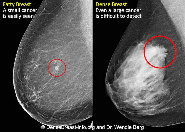
Mammography remains the standard of care in screening for breast cancer and has been proven to reduce the mortality rate [1].  However, in dense breasts, cancers can be hidden/obscured on mammography [2,3] (Fig.1) and may go undetected until they are larger and more likely to present with clinical symptoms [4]. Breast density has also been identified as the most prevalent risk factor for developing breast cancer [5].   

Women with dense breasts are BOTH more likely to develop breast cancer and more likely to have that cancer missed on a mammogram [5]

Fig. 1 – Cancer on a mammogram of a fatty vs a dense breast

What is Dense Breast Tissue?

Breasts are made of fat and glandular tissue, held together by fibrous tissue. The more glandular and fibrous tissue present, the “denser” the breast. Breast density has nothing to do with the way breasts look or feel. Whilst dense breasts are normal and common, dense breast tissue makes it more difficult for radiologists to detect cancer on a mammogram.  

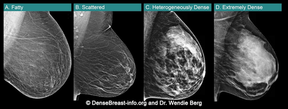
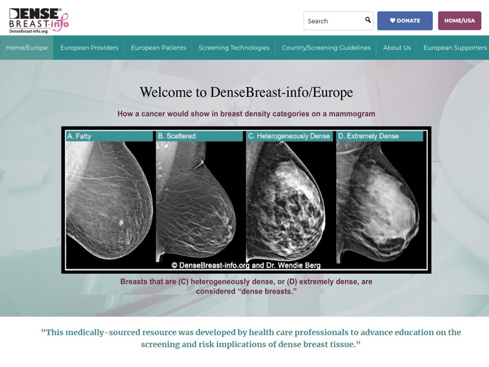
Breast density is determined through a mammogram and described as one of four categories (Fig. 2), (A) Fatty, (B) Scattered, (C) Heterogeneously Dense, (D) Extremely Dense.  Breasts that are (C) heterogeneously dense, or (D) extremely dense are considered “dense breasts”.  Fig. 2

Figure 2

Dense Breasts Facts

  • 40% of women over age 40 have dense breasts.
  • Dense breast tissue is an independent risk factor for the development of breast cancer; the denser the breast, the higher the risk.
  • Mammograms will miss about 40% of cancers in women with extremely dense breasts.
  • Women with extremely dense breasts face an increased risk of late diagnosis of breast cancer.
  • In these women, screening tests, such as ultrasound or MRI, when added to mammography, substantially increase the detection of early-stage breast cancer.

Dense Breast Educational Resources

DenseBreast-info.org/Europe is the world’s leading website about dense breasts. This medically-sourced resource is the collaborative effort of world-renowned experts in breast imaging and medical reviewers. Fig 3.

Figure 3


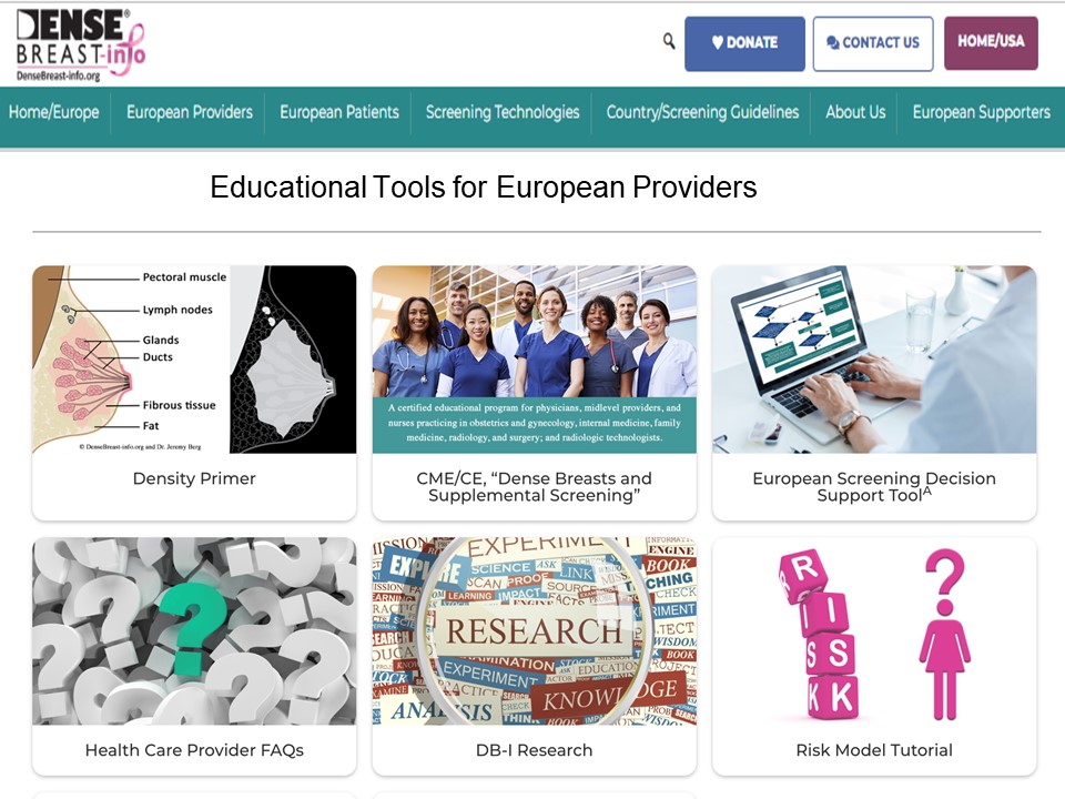
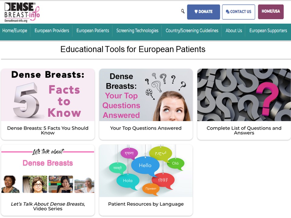
The website features educational tools for both European Patients and Providers Fig. 4. (a and b)

Figure 4 (a)

CME Course – Learn Why Breast Density Matters!

The DenseBreast-info.org resource includes a free CME/CE course, Dense Breasts and Supplemental Screening suitable for primary care healthcare providers, including family medicine, internal medicine, and OB/GYN physicians and midlevel providers, as well as radiologists, and radiologic technologists (UEMS-EACCME® mutual recognition for AMA credits).

A growing number of medical organisations link to the DenseBreast-info.org website, including the EFRS (European Federation of Radiographer Societies) and the Society of Radiographers.  


Figure 4 (b)


The website includes breast screening guidelines in Europe. A comparative analysis table summarises the guidelines in each country.

NHS Breast Screening Programme

Currently in the UK, population routine screening mammograms are offered to women aged 50–74, every 3 years. Though dense breasts affect the likelihood that a cancer will be masked and increases a woman’s risk for developing breast cancer, it is not part of UK data collection. A woman’s breast density is not assessed, not recorded in medical records, nor reported to her. For diagnostic purposes, this may differ. However, in many other European country screening programs, a woman’s breast density is assessed, recorded, and the woman’s personal breast density category is included in the mammography report.

News in Europe:  the EUSOBI Recommendations

Population based breast screening guidelines vary across Europe. In the UK, asymptomatic women attending routine national breast screenings receive mammography alone. In some countries (e.g., Austria, Croatia, Hungary, France, Serbia, Spain, Switzerland) screening guidelines for women with dense breasts include that they be offered supplement ultrasound following a mammogram.

Following recent MRI screening trials there is cumulating evidence which confirms that women with dense breasts are underserved by screening with mammography alone [7,8]. In March 2022, new guidelines were issued in Breast cancer screening in women with extremely dense breasts by the European Society of Breast Imaging (EUSOBI) [9] highlighting the growing evidence, particularly the results of a randomised, multicentre controlled study, the Dense Tissue and Early Breast Neoplasm Screening (DENSE) Trial. [7,8]

The European Society of Breast Imaging 2022 recommendations now step away from the one-size-fits all approach of mammography that is currently adopted by most European screening organizations and advocates for tailored screening programmes. There is compelling evidence that the new recommendations enable an important reduction in breast cancer mortality for these women. 

Summary of the EUSOBI Recommendations

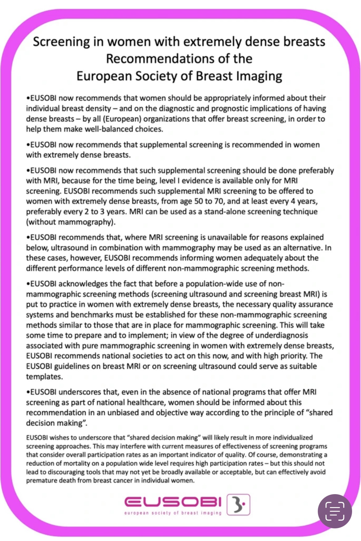
Below is EUSOBI’s summary graphic of the recommendations (Fig. 7) that highlight:   

  • Supplemental screening is recommended for women with extremely dense breasts.  
  • Supplemental screening should be done preferably with MRI …. where MRI is unavailable… ultrasound in combination with mammograph may be used as an alternative.

In addition to recommended additional screening in women with extremely dense breasts, note that EUSOBI recommends that “women should be appropriately informed about their individual breast density in order to help them make well-balanced choices.”

EUSOBI acknowledges that it may take time before the new recommendations are implemented in Europe and that the level of implementation is dependent on the resources that are available locally. 

It is important to emphasize that the EUSOBI recommendations highlighted in this article are not yet guidelines in Europe. Of course, it is hoped that in Europe, national breast screening committees try to implement these recommendations as soon as possible to benefit women.   


Figure 7

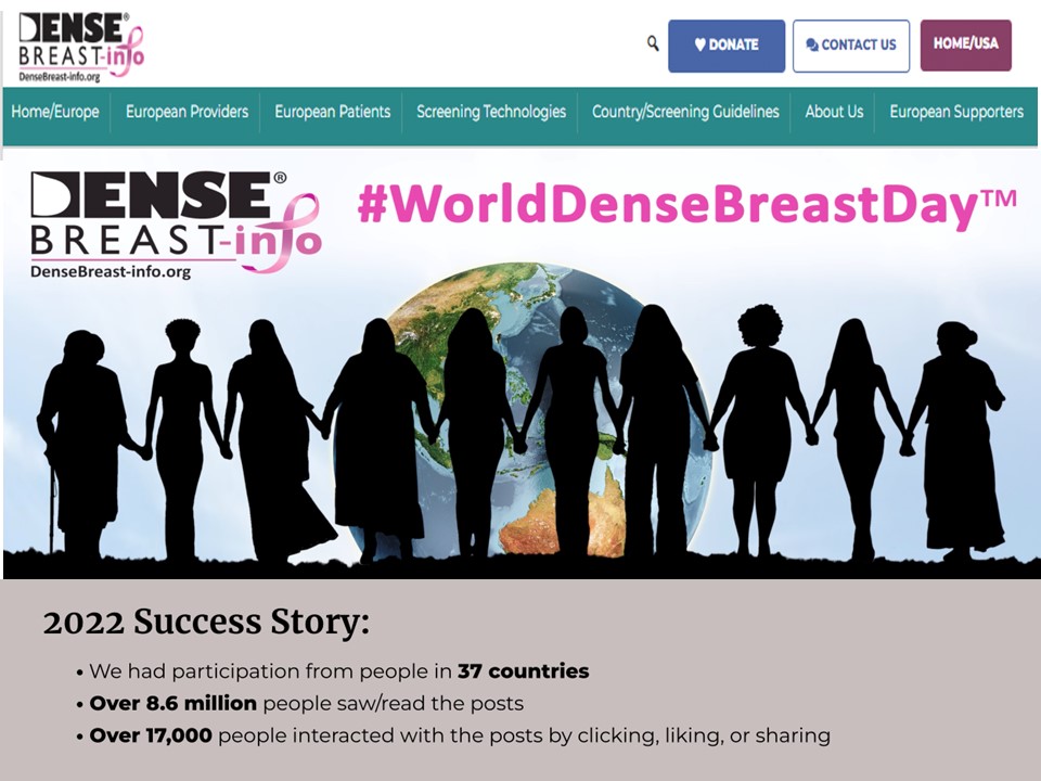
World Dense Breast Day Success!

DenseBreast-info.org launched the first #WorldDenseBreastDay on 28 September 2022.

Nearly 100 posts with great images were created and ran for 24 hours across social media channels.  Analytics detailed participation from people in 37 countries, over 8.6 million people saw/read the posts and over 17,000 people interacted with the posts.

The purpose of the day is to raise awareness about dense breasts and share medically-sourced educational resources available for women and health providers.                                                                                          

Please join us next year for #WorldDenseBreastDay which will take place on 27 September 2023!                               

Take Home Message:

  • Breast density can both hide cancers on a mammogram and increases the risk of developing breast cancer.
  • Women with dense breasts benefit from additional screening tests after their mammogram
  • Breast density education and access to supplemental screening can mean the difference between early- or late-stage diagnosis
  • Physicians should be educated and prepared to have patient conversations about breast density   
  • For more information about Dense Breasts visit: DenseBreast-info.org/Europe

——————————————————————————————————-

1, Tabar L, Vitak B, Chen T H et al. Swedish two-county trial: impact of mammographic screening on breast cancer mortality during 3 decades. Radiology 2011;260:658-63

2. Hooley RJ, Greenberg KL, Stackhouse RM, Geisel JL, Butler RS, Philpotts LE (2012) Screening US in patients with mammographically dense breasts: initial experience with Connecticut Public Act 09-41. Radiology 265:59–69

3. Kolb TM, Lichy J, Newhouse JH (2002) Comparison of the performance of screening mammography, physical examination, and breast US and evaluation of factors that influence them: an analysis of 27,825 patient evaluations. Radiology 225:165–175

4. RoubidouxMA, Bailey JE,Wray LA, HelvieMA(2004) Invasive cancers detected after breast cancer screening yielded a negative result: relationship of mammographic density to tumor prognostic factors. Radiology 230:42–48

5. McCormack VA, dos Santos Silva I (2006) Breast density and parenchymal patterns as markers of breast cancer risk: a metaanalysis. Cancer Epidemiol Biomarkers Prev 15:1159–1169

6. Vourtsis A, Berg W A. Breast density implications and supplemental screening. Eur Radiol 2019;29:1762-77.

7. Bakker M F, de Lange S V, Pijnappel R M et al. Supplemental MRI screening for women with extremely dense breast tissue. N Engl J Med 2019;381:2091-102.

8. Stefanie G. A. VeenhuizenStéphanie V. de LangeMarije F. BakkerRuud M. PijnappelRitse M. MannEvelyn M. MonninkhofMarleen J. Emaus, Petra K. de Koekkoek-Doll Published online: Mar 16 2021 https://doi.org/10.1148/radiol.2021203633Radiology Vol. 299, No. 2 Supplemental Breast MRI for Women with Extremely Dense Breasts: Results of the Second Screening Round of the DENSE Trial

9. Mann, R.M., Athanasiou, A., Baltzer, P.A.T. et al. (2022) Breast cancer screening in women with extremely dense breasts recommendations of the European Society of Breast Imaging (EUSOBI) Eur Radiol 32, 4036–4045 

Cheryl Cruwys is a British breast cancer patient, advocate, author and educator. While living in France (2016) she was diagnosed with early-stage breast cancer and credits the early detection of breast cancer to the French standard practice of performing supplemental screening on dense breast tissue. She is founder of Breast Density Matters UK, European Education Coordinator at DenseBreast-info.org/Europe, a member of the European Society of Radiology Patient Advisory Group and a Patient Rep on the ecancer.org Editorial Board.

Cheryl works at the European level with patient advocacy and medical societies, attends/presents at key scientific symposiums and works with international breast imaging experts to disseminate education on dense breasts. DenseBreast-info.org 

Adapting to a new way of treating

Dr Ben George

Over the last 18 months, GenesisCare has treated more than 170 patients on the UK’s first ViewRay MRIdian MR-linac and adopted SMART planning as a new way of working. Here, Ben George explains why this latest hypofractionated technique has proven to be one of the success stories of the COVID-19 era.

Stereotactic ablative radiotherapy (SABR) is growing in importance in the curative cancer pathway. Increasingly, it offers patients the opportunity to enjoy relatively long periods of disease control where previously they would have been considered for palliative treatments. During COVID-19, the scales have tipped even further in favour of hypofractionated techniques because protocols have been revised to limit the risk of patient infection. More recently, attention has turned to stereotactic ablative MR-guided adaptive radiotherapy (SMART) – the most exciting development in radiotherapy for years, with the potential to treat previously inaccessible targets.

GenesisCare has been the first in the UK to adopt SMART, installing the first ViewRay MRIdian MR-linac just over a year ago. Since then, we have treated over 170 patients, some of which are the most challenging in the world from a radiotherapy perspective, such as pancreatic, central lung and now renal cell carcinomas. MRIdian sits within our SABR offering, which is run by a specialist team of oncologists, physicists, dosimetrists, and radiographers. Over an intensive 18 months, we have adopted a completely new way of working and overcome the challenges of a pandemic to treat patients not just from across the UK, but also from around the world.

SMART explained

The MRIdian MR-linac combines a 0.35 T split superconducting magnet with a 6 MV linear accelerator. This gives it unique advantages over conventional external beam radiotherapy linear accelerators, which rely on kV cone-beam CT (CBCT) imaging, and enables an entirely new approach to treatment.

First, using MRI instead of CBCT provides superior soft-tissue visualisation. This increased imaging capability allows the treatment to be adapted at each fraction based on the daily position of the target and nearby organs at risk (OARs). This is in marked contrast to external beam treatment with CBCT, where anatomy captured in the CBCT is simply rigidly matched against a planning CT. This rigid registration is then used to calculate the movements required to shift the patient into the correct position for treatment.

Second, the MRIdian takes images continuously throughout the treatment period to not only monitor the patient position, but also turn the treatment beam on and off. This is carried out as the patient’s anatomy moves through the breathing cycle.

This combination of enhanced visualisation and real-time imaging adds a layer of certainty in the delivery of treatment.

  • The MRIdian on-table adaptive planning system generates a new, optimised treatment plan for each fraction. This accounts for these day-to-day anatomical variations when the patient is in the treatment position.
  • Treatment delivery is then automatically gated so that the dose is only delivered when the target is in the optimal position. The machine is able to monitor every intrafraction motion caused by breathing or organ-filling.

As a result of these factors, we can design plans which deliver a higher dose, more precisely than with conventional SABR. There is no need for invasive fiducial marker insertion and any uncertainty is removed. Moreover, we can reduce planning target volumes, remove internal target volumes, and minimise the amount of tissue irradiated.

SMART has led to a paradigm shift in how some cancers are treated. In particular, it can benefit cancers in areas where there is significant inter- or intrafraction motion of either the target or OARs. Across the global community, MR-linac centres are now treating novel indications, such as renal, central lung and hepatobiliary tumours, and achieving clinical outcomes not previously thought possible. It is not simply a case of improving on an existing treatment – for some tumour types, SMART is facilitating new referral patterns for patients who may not typically be eligible for radiotherapy.

Pancreatic cancer – a new way of treating

Pancreatic cancer is one such example and of all the tumour sites we are now treating at GenesisCare, this is undoubtedly the one that is breaking most ground, offering new hope for both clinicians and patients.

For decades, surgical resection and adjuvant chemotherapy and radiotherapy have been the cornerstones of primary and secondary hepatobiliary tumours and pancreatic cancer treatment. However, options are limited for many patients. Less than 20% are resectable at diagnosis and not all patients are fit enough for an operation or effective chemotherapy regimens. There is, however, emerging evidence of a dose-response relationship, proving that escalated radiation doses are associated with improved local control as well as overall survival in borderline resectable (BRPC) or locally advanced pancreatic cancer (LAPC). Conventional radiotherapy delivers a comparatively homogenous radiation dose to the target volume. In contrast, SABR treatments combine advanced image guidance systems, accurate dose delivery and hypofractionated regimes. This is to facilitate a deliberate heterogeneous dose distribution across the target. This means the radiation tolerances of surrounding OARs are respected, while the tumour receives a higher, ablative radiation dose. A number of SABR studies have yielded good results in the treatment of large hepatobiliary tumours, with 1-year local control exceeding 90% and acceptable toxicity. Furthermore, delivering these hypofractionated ablative doses of radiation over a shorter treatment schedule has the potential to reduce the burden of treatment on patients.

However, with conventional SABR this therapeutic approach is often limited by concerns regarding organ motion and the possibility of developing small bowel radiation toxicity. As a result, many patients are only being treated with systemic agents. This is a prime example of where the elements of SMART on an MR-linac can facilitate an effective radiation dose escalation, while still respecting the radiation tolerance of normal tissues and surrounding OARs. In fact, using an MR-linac, it has been possible to successfully increase the prescribed dose in patients with primary pancreatic cancer. The previous standard dose was 33 Gy in five fractions, but SMART enables us to escalate the prescribed up to 40 Gy or even 50 Gy in five fractions. At the time of writing, 30 patients have been treated on the MR-linac for pancreatic tumours at GenesisCare.

Compassionate Access

The significance of MR-linac as an innovation in cancer treatment can’t be understated and, although at GenesisCare we are offering it in a private setting, we are committed to sharing the benefits of this technology with the wider medical community. Patients with localised pancreatic cancer have variable access to precision radiotherapy in the UK. The n-SARS-CoV-2 pandemic has further disadvantaged this patient group by reducing the availability and safety of surgery and chemotherapy. Considering this, since 2020 GenesisCare in association with GenesisCare Foundation, UK charity, Pancreatic Cancer Research Fund, ViewRay and University of Oxford have been treating NHS patients with localised pancreatic cancer with SMART at no costThe programme, which is run through a partnership with the University of Oxford, is generating preliminary clinical and patient-reported outcome data on a UK cohort. This will inform the design of subsequent randomised clinical trials and help to embed SMART in UK oncology practice.

A new way of working

With any new technology, there comes a learning curve. MR-linac represents a significant change in working practices. It demands a style of inter-disciplinary working which challenges the norms.

In a standard radiotherapy workflow, a patient will receive a treatment planning CT one to two weeks before the start of treatment. During this time, several steps are carried out by a team of dosimetrists, physicists, doctors and radiographers to produce a treatment plan ready for the patient’s first fraction. These steps include contouring the treatment target and OARs and optimising the machine parameters to deliver the prescribed dose to the target while sparing critical structures. This is followed by reviewing the dose distribution, checking the planning process to ensure no errors have occurred and performing an independent dose calculation.

As part of the on-table adaptive workflow, the time taken for this process must be reduced from days to minutes. In order to achieve this, close inter-disciplinary working between the team is required. The need to undertake a number of complex tasks during each adaptive treatment also increases the time for each fraction to around one hour.

The MRIdian workflow involves a Clinical Oncologist on-site during treatment to oversee the daily adaption. To maintain a treatment schedule at GenesisCare, this has meant that clinicians had to be trained to contour all areas of anatomy, often working outside their main area of specialism. Equally challenging was the need to acquire skills in MRI interpretation, which for some specialities is not routinely used as a diagnostic modality. These were all skills that needed to be honed and validated before any patients could be treated on the MR-linac. In our case, we spent many hours learning with colleagues in MR-linac centres of excellence around the world. Twelve months later, we are experts in this field and have treated over 170 patients.

A body of evidence

There is a growing body of data as the global MR-linac community treats ever more and complex cases. We brought this international best practice to GenesisCare and have treated complex and challenging cases, including central lung, pancreas and reirradiation within our first year. We have many case studies available on our website genesiscare.com/mridian/case-studies. We already knew that the technology could deliver, but it was the confidence in our processes and the ability of our team to implement an adaptive workflow in a time-pressured environment, with a patient on the treatment table, which allowed us to embrace the opportunity that MR-linac presents in radiotherapy.

GenesisCare will install the second MR-linac in the UK in 2021. Through our MagNET programme, we are joining with NHS organisations to support education in the use of MR-guided radiotherapy. Enquiries to: James.Good@genesiscare.co.uk

Dr Ben George, Lead Physicist – MR Linac, GenesisCare

Ben is Lead Physicist – MR Linac at GenesisCare UK. He works as part of a multi-disciplinary team which has established a successful and world-leading SABR service delivering complex MR-guided adapted treatments. He has a PhD in Physics with a strong background in computer science, research and clinical computing. He has over ten years of experience as a Clinical Scientist specialising in radiotherapy in both the NHS and the private sector, and as a research scientist for the University of Oxford.

A podcast radiating positivity

Angela Young explains how the process of making a podcast helped not only others with a diagnosed brain tumour but gave comfort and support to herself as she embarked on a course of radiotherapy.

A brain tumour diagnosis, like all major events, can set in place a chain of emotions, among them anger, fear and denial. It can also make you adjust your priorities in life. I went through all this in 2015 when I discovered I had a Grade 1 benign posterior fossa meningioma. A resection at Addenbrooke’s Hospital in Cambridge was very successful, leaving only a 3mm residuum.

I had been having regular follow up scans, and in 2019, it was thought the growth was significant enough to consider radiotherapy. After the initial shock, I realised that, if successful, it would prevent the cells from growing again and remove the need for annual scans with the associated “scanxiety”. My decision to go ahead now rather than wait for symptoms to appear was influenced by the consultant radiologist Dr Sarah Jefferies who said the benefit of doing so now was that I was “young and fit”, a nice thing to hear at the age of 59.

As a journalist and podcast maker, I am used to getting to grips with a variety of subjects quickly in order to explain them to others. It dawned on me that if I could tell the story of my own treatment, it would give me a sense of control over a process in which one can easily feel helpless. It might also provide information and some light relief to other people going through something similar and their families. The radiotherapy process would be the same for people undergoing treatment for a variety of conditions, not just brain tumours, and so creating a podcast on this topic could reach and potentially help a large audience.

I am very optimistic by nature and I like to see the funny side of things. I believe that if you look closely, you can find humour in most situations. Consequently, I decided the title of the podcast should be “A Sense of Tumour”. I started recording everything that happened, whether by phone call (I had got all the kit I needed for doing this when lockdown started) or recording my own commentary during appointments and tests and arranging interviews, either face to face (with masks on) or via an audio recording platform.

People find podcasts in a variety of ways. One of those is to have a well-known personality or influencer or support group post about them. It helps if you can interview a celebrity or two who will do this. When I asked Victoria Derbyshire (via a mutual friend) if she would talk to me about documenting her very public battle against breast cancer, I had no idea she would later be taking part in the TV programme “I’m A Celebrity, Get Me Out Of Here!”. Victoria appeared in Episode 1 and set the interview bar quite high. Luckily, the Brain Tumour Charity had come on board by this stage and offered to put me in touch with TV presenter Nicki Chapman, who had had a matching meningioma to mine removed last year. She readily agreed to be interviewed and candidly shared the highs and lows she experienced when going through treatment herself. For the final episode, I thought I would chance my luck and ask to speak to Tony Iommi, lead guitarist and song writer with Black Sabbath. He had had radiotherapy a few years ago and embraced some alternative therapies which I wanted to hear about. To my delight, he was more than willing to talk.

The series was meant to inform as well as entertain so I spoke to the medical professionals whom I was meeting and also those at the cutting edge of research into treatment. I interviewed the Chair of Cancer Research UK, Sir Leszek Borysiewicz, about funding for brain tumours. I also had conversations with the “Distinguished Scientist” from Elekta, one of the companies which makes the linear accelerator machine (not a bad job title!) and to many people from the team at Addenbrooke’s, including a medical physicist and a research radiographer. I learned a lot and I hoped that sharing these conversations would also help listeners to understand some of the more complicated parts of the treatment and process more easily.

Bringing the podcast’s listeners on my journey was supposed to feel personal too. I recorded as much as I could at every stage, including the baseline neurological assessment. This is an IQ-style test carried out before the start of a course of radiotherapy to the brain so that if there is any concern about future cognitive function, there is a baseline against which to compare it. One part of the test included listing as many words as possible beginning with the letter F; you can imagine what came to mind! When that episode was released, listeners I came across would shout out words beginning with F to me.

All the way through the treatment I was thinking how I would represent things aurally, such as the MRI machine. These make a variety of loud noises but would wreck any recording device in the vicinity. When I managed to open my eyes under the thermoplastic mask which holds the head in place on the linear accelerator, part of the machine going over me looked like a spaceship. Friends and family had each contributed a song for my radiotherapy playlist; that day the song was Mr Blue Sky and it had got to the instrumental part, which made me think of a science fiction movie. I was working out how to recreate the impression for the podcast. Thinking about this during the session took my mind off what was going on.

By the time you read this, I will have finished the treatment and will be waiting for a scan to see how successful it has been. I am, of course, hoping for the best. I would also like to think that the podcast series has been useful to patients and their families, to radiotherapists, to manufacturers and anyone else involved in this fascinating process. I also hope that it inspires anyone looking for a positive and creative way of dealing with a diagnosis of any kind to take control of what they can, focus on something meaningful and use their good days to bring strength to others. After all, positivity radiates.

Listen to “A Sense of Tumour” here

About Angela Young

Angela Young founded Cambridge Podcasts in 2018 to help clients showcase their expertise and establish themselves as the go-to person in their field. She is a former BBC radio journalist who has worked as a reporter, producer, news reader and news editor. She has taught law and journalism at the BBC and media handling at the prestigious Institute for Management Development in Lausanne. She studied law at Cambridge as a mature student and has lived in the city for 28 years. www.cambridgepodcasts.co.uk info@cambridgepodcasts.co.uk

“The Radiology Reset Button – overcoming the normalcy bias”

Fodi KyriakosFodi Kyriakos explores how the COVID-19 pandemic could be the catalyst for change in radiology and encourages our community to grasp the opportunity to “seize the moment”  and plan for recovery.

At the beginning of 2020, if someone had told radiology leaders that all NHS outstanding reporting backlogs would be reduced to virtually zero by May, I’m sure they would have looked at you in disbelief and asked what sorcery had been involved, but this situation is exactly where we find ourselves today.


Normalcy Bias – Noun [edit]

normalcy bias (plural normalcy biases)

The phenomenon of disbelieving one’s situation when faced with grave and imminent danger and/or catastrophe. As in over focusing on the actual phenomenon instead of taking evasive action, a state of paralysis.

Historical challenges

In the past, it has often taken lots of effort to either invoke or accept change of any kind in radiology and for those managing services, there’s also been a certain amount of risk associated with putting your head above the parapet or being a trailblazer. It has been sometimes easier to follow the well-trodden path rather than to create a new one. Workloads and budgetary constraints have also been a disabler, restricting decision making to the ‘here and now’. This has resulted in failing, or in most cases, not being able to foresee or plan for events that have never happened before, such as an event like a pandemic crisis. Psychology refers to this state of being as normalcy bias. For those who are not familiar with the term, you will certainly be aware of its connotations and radiology now finds itself at this cross-roads.

Ever since the introduction of digital radiography and PACS, NHS radiology reporting backlogs have been a contentious issue among experts, and a recurring feature in the mainstream media! Often being highlighted (and with some justification) in relation to areas such as missed cancer diagnosis, where even the slightest of delays can have a significant bearing on the overall outcome.

Serious backlogs

The extent to which backlogs were a serious issue in the UK was further exacerbated by various Care Quality Commission (CQC)  inspections, which raised concerns regarding reporting backlogs that resulted in delayed or missed diagnosis of conditions that may have otherwise been picked up.

By the end of February 2020, the situation of backlogs was as much an issue as at any time before. Insufficient reporting capacity had led to a build-up of outstanding reports, which in turn meant that outsourcing was at its highest ever levels and growing pressures to meet new deadlines, such as the cancer pathway targets, were increasingly exposing the lack of options available to resolve the problem.

So, you would have been excused if you thought that a crisis such as the COVID-19 pandemic would simply exacerbate the reporting challenges facing radiology. However, this has not been the case. Instead, we have witnessed radiology’s own “clear the decks” exercise, where in fact the complete opposite situation has occurred, resulting in backlogs across the UK being virtually eliminated. Who would have thought that the worst crisis to hit the country (and the world) in 75 years would be a catalyst for NHS radiology departments to press the reset button?

Reset image

Of course, we recognise the superficial nature of this situation. During the pandemic, practically all routine referral activity came to a grinding halt, which allowed radiology to concentrate on COVID-19 and Emergency Department (ED) patients. Chest X-rays and CTs were identified as two of the key diagnostic tools for the virus, but the volumes were manageable. Accident and Emergency footfall was reduced to almost 50% of its usual figures, so reporters were practically able to deliver a ‘Hot-Reporting’ examination for every patient requiring imaging. Something which ED and Intensive care unit (ICU) consultants have grown quickly accustomed to.

During this time, radiology was also still required to work to critical staffing levels, so radiographers and radiologists were covering 24/7 rotas, but due to the lack of activity outside of portable X-ray scanning in ICU, many staff were not being utilised. So, while this enabled the catch up in radiology reporting to take place, what we witnessed was the ‘ying and yang’ of radiology. On the one hand, integral to the continuity of a patient’s pathway and critical to defining an outcome – AND on the other hand, completely dependent on throughput from referrers to maintain activity levels.

Seizing the moment!

So what happens next? Well, in a world where we can guarantee almost nothing, in this situation, we can guarantee that radiology will remain the centre point for the recovery phase of the pandemic, but with the added challenge of complying to ‘social distancing’ and ‘equipment cleaning’ guidelines, how do we manage the continuation of treating COVID-19 patients, while reintroducing ‘business as usual’ and ‘deferred’ patients whose treatment has been delayed?

The “Reset Button” has enabled something else to happen. For the first time, there is now some headspace to plan for the recovery phase and for the next phase at least, there is now funding available to support the recovery. So how do we avoid going back to where we were before the pandemic? How do we seize the moment?

Time to make the changes!

Albert Einstein once famously said: “We can’t solve problems by using the same kind of thinking we used when we created them.” This quote has never been more poignant in the present day and while the pressure to manage change will be at its highest, this is the right time to make these changes happen! With the benefit of ‘The Reset Button’, if we can learn from the past and apply new ways of working moving forward, we can avoid falling into the trap of the normalcy bias and witness the radiology reset button offering a new, efficient and more streamlined radiology department moving forward.

Everything you wanted to know about radiology but were afraid to ask…

On Wednesday 17 June, a live event organised by InHealth, in partnership with The British Institute of Radiology and the Society of Radiographers is taking place, titled: “The Radiology reset button has been pressed”. The aim is to tackle these challenges and support radiology managers as they enter the recovery phase. It will bring together senior figures from radiology and within healthcare to offer insights, opinions and advice on how we can approach this coming period and use what positives we have experienced during the pandemic to create service improvements throughout radiology.

There will be opportunities for radiology managers, clinical leads, radiographers and radiologists to put their questions to the speakers in the panel discussions after their presentations.

REGISTER FOR THE RADIOLOGY RESET BUTTON HAS BEEN PRESSED HERE

(The event is free for all)
About Fodi Kyriakos

Mr Fodi Kyriakos is a former director of RIG Healthcare and founder of RIG Reporting,
the UK’s first provider of external radiographer reporting services. In 2016 he joined The InHealth Group following its acquisition of RIG Reporting and is now the Head of Reporting across the Group. His service specialises in delivering plain film reporting solutions and is the only provider to offer both on-site and telereporting services.
Fodi has over 22 years experience in workforce and staffing solutions and 17 years working exclusive within Imaging and Oncology. He is a member of the Institute of Healthcare Managers and a regular contributor of professional development events across radiology.


Bringing together Science, Faith and Cancer Care

Slide2

The Revd. Canon Dr. Mike Kirby, Chair of the BIR Oncology and Radiotherapy Special Interest Group, has a wealth of experience as a senior radiotherapy physicist, working on national guidance, developing clinical practice and teaching radiography students. As if this doesn’t keep him busy enough he has also taken on the role of Canon Scientist at Liverpool Cathedral where he is working to encourage dialogue and discussion about science and faith. Here he explains what the role involves.

I began work in the UK’s National Health Service more than 30 years ago, as a Radiotherapy Physicist at the Christie Hospital, Manchester UK.  Alongside my routine clinical work, my main research interest was in electronic portal imaging and portal dosimetry.  I then helped set up Rosemere Cancer Centre in Preston, UK from 1996 as deputy Head of Radiotherapy Physics and Consultant Clinical Scientist there.  During that time I contributed to and edited national guidance documents such as IPEM Reports 92, 93 and 94 and the multidisciplinary work, ‘On-target’.

My work moved back to the Christie in 2007 and as Head of Radiotherapy Physics and Consultant Clinical Scientist for the Satellite Centres, I helped to lead their development in Oldham and Salford as part of the Christie Network. My research and development work has primarily focused on electronic portal imaging, developing clinical practice and equipment development.

Mike Kirby4

More recently my focus has been on teaching and learning for radiotherapy education as a lecturer (Radiotherapy Physics), especially using VERT, for Radiotherapy programmes in the School of Health Sciences, Liverpool University; but always with a focus on the wider picture of radiotherapy development having served on both IPEM and BIR committees throughout my whole professional career.


Alongside my scientific work, I am a priest in the Church of England; having trained and studied at Westcott House and the Universities of Cambridge and Cumbria, I hold graduate and postgraduate degrees in Theology.

Mike Kirby

My ministry has mainly been in the Cathedrals of Blackburn, Chester, and Liverpool (Anglican) where I was Cathedral Chaplain.  I have recently (Feb 2020) become a Residentiary Canon of Liverpool Cathedral, with the title of Canon Scientist the primary aim of which is to encourage dialogue and discussion about science and faith.

 I am a member of the Society of Ordained Scientists and have given numerous talks on Science and Faith to schools, colleges, churches and other institutions.  These have included organising lecture series with world renowned speakers at Blackburn (2016) and Chester (2018) cathedrals; a third series was delivered at Liverpool Cathedral in May 2019, and a fourth series is planned for May 2020.

My role is to consider all sciences (physical, clinical, social) in ecumenical and multi-faith environments.  So I will look to work with initiatives already developing in other Christian traditions, other faiths and secular organisations to discuss current challenges, such as climate change, medical ethics, health initiatives and information for cancer, dementia and mental health issues etc..

Mike Kirby2.jpg

My work will be part of the clear faith objectives of the cathedral as a place of encounter for everyone, through events and initiatives within the cathedral, but also beyond.  This will include services focusing on health issues and pastoral challenges (such as bereavement and loss); events engaging with science, its wonders and challenges; fostering further relationships with local and wider communities on science and healthcare education, and with academic and scientific institutions too; encouraging scientific and ethical engagement with schools and colleges, as I have done so previously in both Chester and Blackburn dioceses.

I will be encouraging Christians and Christian leaders to understand science and engage with it more, alongside other national projects such as the recently announced ECLAS (Engaging Christian Leaders in an Age of Science) project of Durham and York universities and the Church of England.  As a self-supporting minister (one whose paid employment is outside of the church), I will also look to encourage and highlight the tireless work of many others who already do this within the diocese and the wider national church.

Within all of this, I have always seen my vocation as being one within God’s service, for all people, with my work for cancer patients being right at the heart of it.

006

If you have any questions for Mike, you can send him an email at sigs@bir.org.uk

Mike is the co-author of the international student textbook on On-treatment Verification Imaging: a Study Guide for IGRT, through CRC press/Taylor and Francis with Kerrie-Anne Calder. They are both contributors to the updated UK national guidance on IGRT due out in 2020.

Mike, with the support of the SIG, has helped to organise a range of events for radiographers, physicists, dosimetrists, radiologists and oncologists. See the full programme here


Help! I’m (not) a leader, get me out of here!

Elizabeth Loney

Do you ever wonder how you got where you are? Are you sure you see yourself as others do? 

Dr Elizabeth Loney, Consultant Radiologist and Associate Medical Director,  reflects on imposter syndrome and offers tips on how to manage it.


How many times have you sat in a meeting and looked around the room thinking, “what on earth am I doing here? Everyone else knows way more about this than I do, and they know it!”

The first senior management meeting that I attended started with reviewing the minutes of the last. As I read through the document, I realised I had no idea what much of it said—death by TLAs (three letter acronyms!). I nudged the person next to me and said, “what does … stand for?” They shrugged their shoulders and whispered to the person on their other side “what does … mean?” It took five people down the line before someone knew what it was! I found that reassuring, but also slightly scary. The fact that other people were in the same boat made me feel less like an idiot, but at the same time, how could such a senior group not understand the jargon and why had they said nothing? So… lesson one: be curious and not afraid to ask questions. You’re probably just asking what most people are thinking anyway!

About six months ago I started the Nye Bevan Programme with the NHS Leadership Academy. If I pass, I will allegedly have proven myself ready for an NHS executive leadership role. There are around 48 others in my cohort, all senior leaders in different areas of the NHS. What the heck am I doing there?! I’m just a doctor, not a leader. I might sort things out for people as Clinical or Divisional Director but I’ve never felt more like a “public servant” than when in a “leadership role”. I had serious Imposter Syndrome. The first residential was entitled “Knowing Yourself and Others” and was all about the impact you have on others as a leader and why you act as you do—unconscious bias and all. It was a traumatic experience for me. I did so much “reflecting” I felt like a mirror! I couldn’t do it—just give me a few scans to report! I’m not a leader—get me out of here. However, I got chatting to others that week and realised that pretty much everyone else in the room felt the same. Most people suffer with this issue at some time—and if you don’t, why not? A little humility is a wonderful thing.

Are you affected by low self-confidence? At times like this, seeking peer feedback can be helpful. As part of the course I had to send out a questionnaire asking others I had led on a work programme for anonymous feedback. That was scary! I asked questions including “what do I do well?” and “what could I do better?” I half expected to be slated but, to my surprise, the feedback was really positive. My view of myself was distorted. I may not see myself as a leader but apparently others do! So… lesson two: when you feel like an imposter remember that many others in the room feel the same way. There must be a reason why you are there. What do others see in you, that you do not? What is your role in the group? ‘If not you… who?’

So ends my first blog as Chair of the BIR Leadership and Management SIG… another position I find myself in wondering how I got here! What do I know about leadership? I’m not an expert. However, I do have a passion for self-improvement and a curious nature. Why not join me on my journey to “managerial enlightenment”? We have such a lot to learn from one another.

I hope to meet you in person at the BIR Annual Congress where we will gain inspiration from excellent speakers covering topics on “practical” and “personal” management, including an interactive session by Philips based on their “Insights” programme—expect to be up on your feet! We are also holding our first annual event on leadership, “Leadership 2020” on 31 January 2020. Come along and join us for more opportunities to learn, network and ask questions.

See you there!

Dr Elizabeth Loney,

Chair of the BIR Leadership and Management Special Interest Group

BIR LEADERSHIP 2020 event 31 January 2020

More about the BIR leadership and management SIG here 

Join the open SIG here  (BIR member only)

About Dr Elizabeth Loney

Dr Elizabeth Loney is Chair of the BIR Leadership and Management Special Interest Group (SIG). She is a Consultant Radiologist and Associate Medical Director and Consultant Radiologist at Calderdale and Huddersfield NHS Foundation Trust.


Radiotherapy: 40 years from tracing paper to tomotherapy

NHS

Physicist Andy Moloney and Clinical Oncologist David Morgan reflect on how radiotherapy developed since their early careers


We first met in the autumn of 1981, when the NHS was, at 33 years from its inception, but a youngster. Andy had recently joined the Radiotherapy Physics staff at Nottingham General Hospital after graduating in Physics from the University of Nottingham, and David was returning to the clinical Department of Radiotherapy and Oncology after a year’s Fellowship at the Institut Gustave-Roussy in France. A firm friendship rapidly developed, one that continues to this day.

On reflection, joining the radiotherapy fraternity at that time was a leap of faith. The perceived wisdom amongst many of our scientific and clinical colleagues at the time was that this treatment technique was outdated and overshadowed by radical surgical procedures, new chemotherapy agents and biological modifiers poised to reduce radiotherapy to the history books.

picture 063
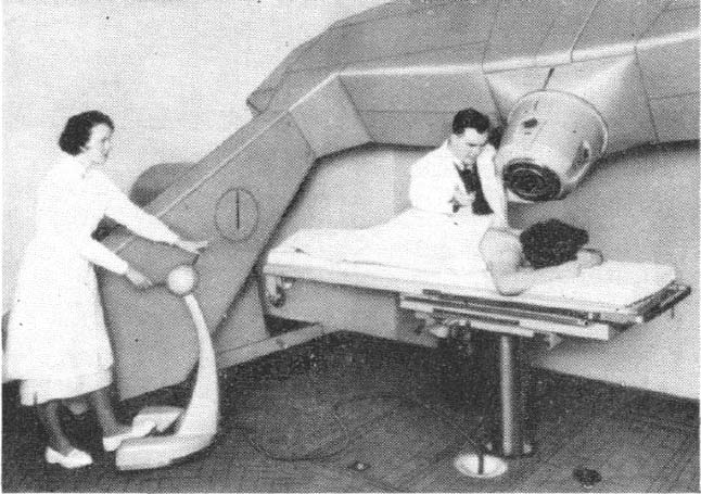
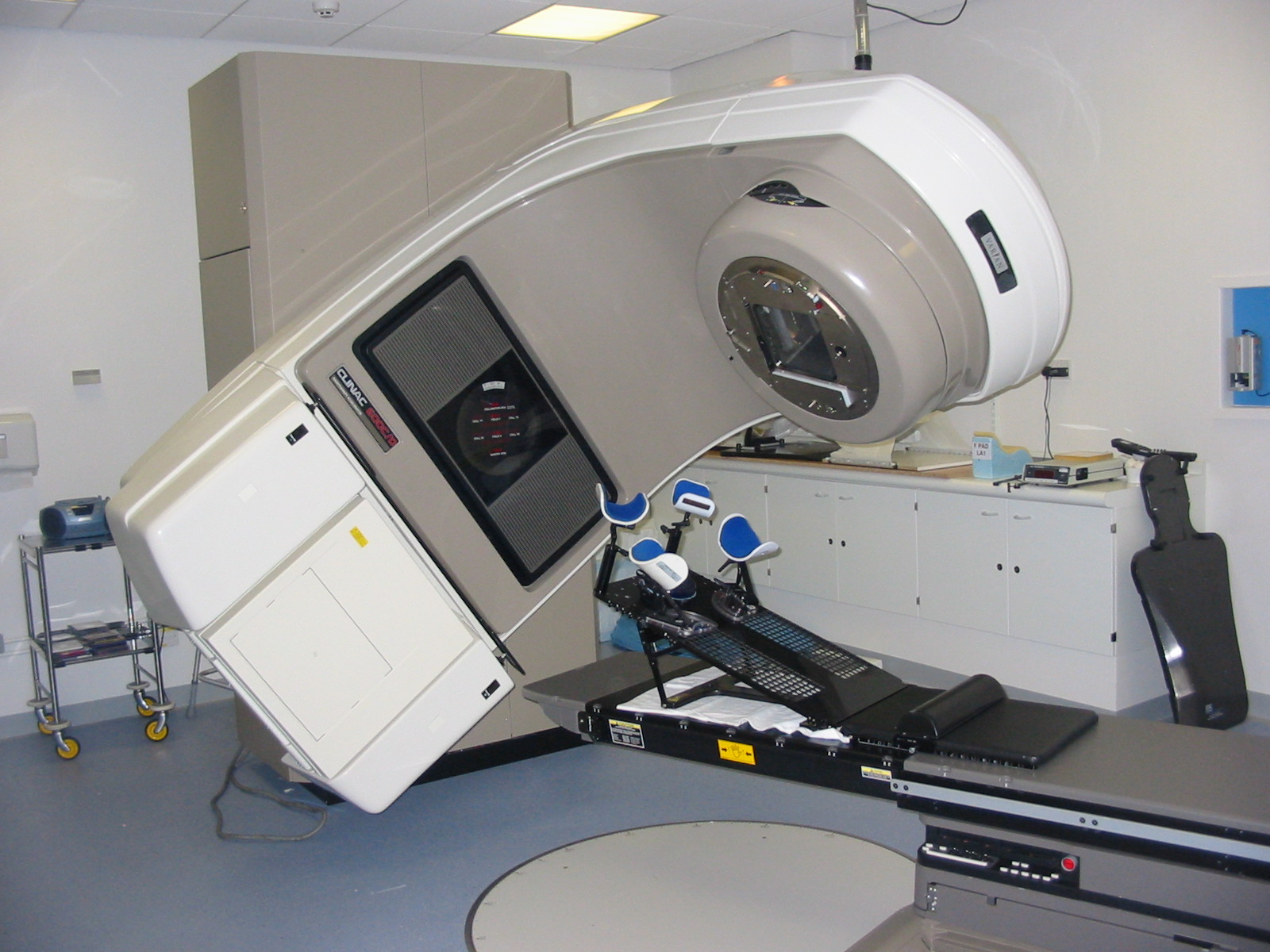
This was a time when, in this Cinderella of specialties, physics planning was achieved by the superposition of two dimensional radiation plots (isodoses) ,using tracing paper and pencils, to produce summated maps of the distribution. The crude patient outlines were derived from laborious isocentric distance measurements augmented by the essential “flexicurve”. The whole planning process was slow and labour intensive fraught with errors and ridiculed by colleagues in the perceived prestigious scientific and clinical disciplines. The principal platform for external beam radiotherapy delivery, the Linear Accelerator (LinAc), had also reached something of a plateau of development, albeit with improved reliability, but few fundamental changes. Caesium tubes were transported from the “radium safe”, locked in an underground vault, to the operating theatre in a lead-lined trolley, where they were only loaded into “central tubes” and “ovoids” after the examination under anaesthetic (which was performed with the patient in the knee-chest position); they were then manually placed into the patient, who went to be nursed on an open ward, albeit behind strategically placed lead barriers.

For no sites outside the cranium was Computer Tomography (CT) scanning available. Magnetic Resonance Imaging (MRI) was still a vision seen only by a small number of enthusiasts.

All these limitations were met by a developing team of scientific and clinical enthusiasts believing in the future of radiotherapy if only technology could deliver solutions to address an improving understanding of the differing cancers and their radiobiology.

picture 066
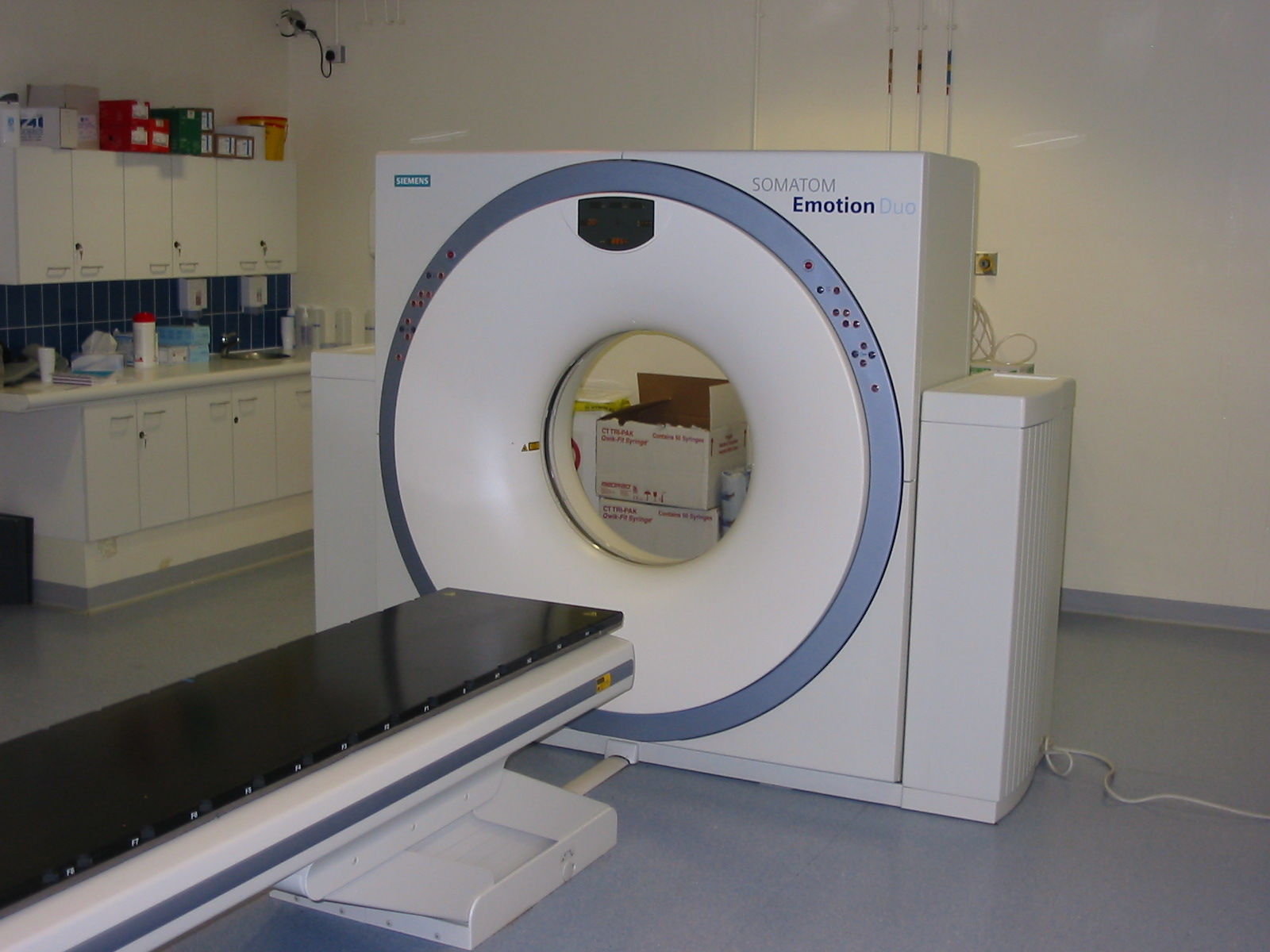
In the latter half of the eighties these solutions began to crystallise. Computers were being introduced across the NHS and their impact was not lost in radiotherapy. Pads of tracing paper were replaced with the first generation of planning computers. The simple “Bentley-Milan” algorithms could account for patient outlines accurately and speedily and optimising different beam configurations became practical. Consideration of Organs at Risk, as defined by the various International Commission on Radiation Units (ICRU) publications, became increasingly relevant. Recognition of the importance of delineating the target volumes and protecting normal tissue required improved imaging and this was provided by the new generation of CT scanners. In the nineties these were shared facilities with diagnostic radiology departments. However, the improvements provided by this imaging, enabling accurate 3-dimensional mapping of the disease with adjacent normal tissues and organs at risk, dictated their inclusion into every radiotherapy department soon after the millennium. The added bonus of using the grey scale pixel information, or Hounsfield numbers, to calculate accurate radiation transport distributions soon followed when the mathematical and computer technology caught up with the task. The value of MR and Positron Emission Tomography (PET) imaging was also recognised in the diagnosis, staging and planning of radiotherapy and the new century saw all of these new technologies embedded within the department.

Mould room technology was also improving with “instant” thermoplastic immobilisation shells replacing the uncomfortable plaster and vacuum forming methods. Custom shielding with low melting high density alloys was becoming routine and it was not long before these techniques were married with the emerging CT planning to provide “conformal” treatments.

picture 067
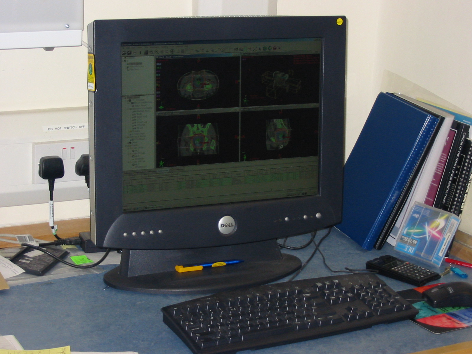
LinAc technology also received added impetus. Computers were firstly coupled as a front end to conventional LinAcs as a safety interface to reduce the potential for “pilot error”. Their values were soon recognised by the manufacturers and were increasingly integrated into the machine, monitoring performance digitally and driving the new developments of Multi Leaf Collimators (MLC) and On Board Imaging (OBI).

The dominos for the radiotherapy renaissance were stacked up, but it needed the radiographers, clinicians and scientists to decide on the direction of travel. Computer power coupled with advanced electro-mechanical design had transformed MLC efficiency and resolution. Conventional conformal planning was now progressively superseded by sophisticated planning algorithms using merged CT and MR images. Intensity Modulated RadioTherapy (IMRT) had arrived in its evolving guises of multiple fixed field, dynamic arc therapy (RapidArc) or Tomotherapy. Whichever technique, they all offered the radiotherapy “Holy Grail” of providing three dimensional homogeneous dose distributions conformed to the Planning Target Volume (PTV) whilst achieving the required dose constraints for organs at risk and normal tissue preservation.

The tools had arrived, but an infrastructure to introduce these “toys” safely into a complex clinical background had also developed alongside. Quality standards (ISO9000), Clinical Trials, Multi Disciplinary Teams and Peer Review were governance mandates for all oncology departments and radiotherapy was leading the way. In forty years, radiotherapy had lost the “Cinderella” image and had been invited back to the clinical ball. Noticeably, breast and prostate adenocarcinoma constituted half of the radical workload.

The question remains of how and why did this transformation occur? Obviously the developing computer power and technology were the pre-requisites for many of the developments, but a key catalyst was the foresight of all of the radiotherapy family from which enduring friendships have been forged. The working lives of the clinicians and physicists involved in radiotherapy planning have probably changed more dramatically than those of any other medical and paramedical groups over the last 35 years.

We may have retired, but we still cogitate about the future direction and science behind this developing and essential cancer treatment and look forward to our younger colleagues enjoying their careers as much as we enjoyed ours.


About David Morgan

david morgan
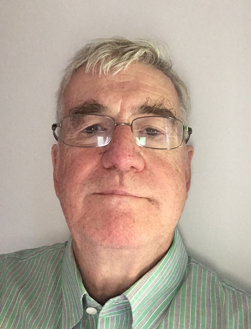
Dr David A L Morgan began training in Radiotherapy & Oncology as a Registrar in 1977, and in 1982 was appointed a Consultant in the specialty in Nottingham, continuing to work there until his retirement in 2011. He joined the BIR in 1980 and at times served as Chair of its Oncology Committee and a Member of Council. He was elected Fellow of the BIR in 2007. He is author or co-author of over 100 peer-reviewed papers on various aspects of Oncology and Radiobiology.


About Andrew Moloney

andy moloneyAndy Moloney completed his degree in Physics at Nottingham University in 1980 before joining the Medical Physics department at the Queens Medical Centre in the same city. After one year’s basic training in evoked potentials and nuclear medicine, he moved to the General Hospital in Nottingham to pursue a career in Radiotherapy Physics and achieved qualification in 1985. Subsequently, Andy moved to the new radiotherapy department at the City Hospital, Nottingham, where he progressed up the career ladder until his promotion as the new head of Radiotherapy Physics at the North Staffordshire Royal Infirmary in Stoke-on-Trent. Over the next twenty years Andy has acted as Clinical Director for the oncology department and served on the Radiation Physics and Oncology Committees at the BIR and was appointed a Fellow in 2007. He has been the author and co-author of multiple peer reviewed articles over the years prior to his retirement in 2017.


When medical physicists wore white coats

NHS

Dr Edwin Aird shares his memories of the revolution he has experienced working in the medical physics department.


Edwin Aird portrait

While I was an undergraduate at Newcastle University (1962), the 2nd year honours group was invited to visit the Medical Physics Department at Newcastle General Hospital. I was so impressed with the department and the range of things they were doing and the application of physics to medicine that I wrote to Professor Frank Farmer (Head of Department at that time) to ask about working there. He responded with a proposal that I apply for a special research grant that he was hoping for locally and that I write again after my graduation.

The following year I got in touch with Frank Farmer again and was accepted on an MSc grant to study “In-homogeneities in Radiotherapy”; one year in the first instance (on a grant of about £700).

Meeting 1970 1

I can’t now remember my first day exactly; not sure what office I had, but I do remember the Professor’s insistence on donning a white coat (provided and laundered by the hospital) when arriving at work, This ‘uniform’ was thought to be a vital sign to patients that physicists were part of the ‘clinical’ team.

I divided my time in that first year between research and clinical work (interestingly, this was part of the philosophy of some of the early physicists, many of whom in the 1930s and 40s had transferred from academic physics, including Frank Farmer); the situation now couldn’t be more different.

Fig 1

Image 1 The gantry mounted linear accelerator at Newcastle General Hospital [ref 2]

So, on the routine side, I began to learn about radiotherapy: its planning and treatment. Newcastle at that time had two cobalt machines (Mobaltron-60s) and two linear accelerators as well as two Marconi 250kV sets and a Philips SXT (Superficial X-rays). This latter set I used extensively a little later in my career to optimise the characteristics of the Farmer chamber (image 1).

The main linear accelerator was manufactured by Mullard (a branch of Philips), which Frank Farmer had installed as the first gantry-mounted 4MV linac (image 2). There were a few stories about the installation of this linac. In particular I remember this: the team had reached the point where they needed to produce X-rays so needed a high atomic number transmission target. Someone found a sovereign, but forgot the amount of heat produced when the high energy electrons hit it. The sovereign promptly melted and fell out of the linac head. (This linac went on to perform 25 years’ service).

Fig 2

Fig 2 Newcastle Simulator (Frank Farmer at Controls) [ref 3]

For those physicists who find quality assurance (QA) on linear accelerators today a huge burden, my recollection of the checks we performed on this linac amounted to: dose measurements and field size checks. There was little attempt to measure flatness since the accelerated electron beam wasn’t bent before striking the target, so no perturbations in intensity across the field were expected.


Fig 3

Fig 3 A Head and Neck Xeroradiograph showing potential. Treatment volume [ref 3]


Treatment planning was done by hand, often mainly using % depth dose, but also with some isodose curves for more complex plans on tracing paper; on to an outline of the patient performed using lead wire and other devices, e.g. callipers. Newcastle was unique in having developed a home-made simulator (using a radiotherapy SXT unit mounted on a gantry (image 2) that allowed an image to be viewed on a Xerox plate immediately without the need for film development (ref 3, see also image 3).

The first computer in the department, which was to revolutionise treatment planning, came to Newcastle in the early 1970s and was called a PDP 8, the ‘Rad8 system’ developed by Bentley and Milan. (image 4 [ref 4]).

Radium tubes and needles were still extensively used at this time for intracavitary and interstitial brachytherapy. I was also required to calculate the dwell times for gynae radium insertions (using ovoids and central vaginal sources with a modified Manchester system for the gynae and radium needles, mainly in back of tongue with radiographs and Paterson Parker tables). Following the calculation of the radium dwell times I would then go up to the ward to discuss the removal times, for each patient, with sister on the ward. Amazing now to think how much radioactivity (up to 105mg – equivalent to approximately 389 MBq-or more) for gynae insertions was handled by several different groups of staff at that time.

In those early years I also learnt the elements of radiation protection and nuclear medicine. Radiation Protection (guided by a ‘Code of Practice’; an excellent document that was used to help write the ‘Guidance Note’ now used under Ionising radiation Legislation) was regionally organised. Very early in my career (there were only two of us to perform the external radiation work: myself and MJ Day), I found myself organising visits within Northumberland, Cumbria and parts of Durham. (See The Regional Centre below). These were more as inspections than to make many measurements (not the very extensive QA that is performed now, although we did use film or image intensification – only recently developed – to look for faults in lead aprons; and we measured exposure levels in and out of beam with the ‘37D’ (Pitman) dosemeter (originally developed by Sidney Osborne as an excellent versatile instrument for exposure measurements ref 5). I learnt a pattern of inspection of barriers, filtration, lead aprons etc. very quickly. [In the early 1970s the NRPB decided to do their own inspections and were surprised to find that, because the HPA had organised things so well there was little need for NRPB to get too heavily involved].

My memories of nuclear medicine: incredibly slow rectilinear scanners, prior to the commercial development of gamma cameras; kidney function (renogram) using two ‘D’-shaped detectors (scintillation) with manual optimisation of their positions connected to count rate meters and chart recorders.

The regional centre

I’m not sure how many hospitals this covered in my early days in Northumberland, Tyneside, Durham and Cumbria. I know when Keith Boddy implemented Frank Farmer’s plan to have a small physics department (where gamma cameras were installed in district general hospitals) there were thirteen hospitals with physics departments in the region. Prior to this – from about my 3rd year at Newcastle – I was delegated to look after the small centre in Carlisle where there were two Marconi 250kV sets, an SXT and some iodine treatment. In the 1950s this had been the job of Jack Fowler who used some of his time to study arc therapy on 250kV (see ref 6).

Rad 2014 photo

Edwin Aird (right) receiving the BIR Sylvanus Thompson Award from Andy Beavis

In those early years I also found time to help develop differential X-ray absorptiometry to measure antimony in the local Tyneside Antimony workers’ lungs (organised by the Newcastle University Department of Industrial Health). I was also able to build on this experience to develop my own equipment, using characteristic X-rays, to measure bone mineral in the femur. This clinical work allowed me to meet a new set of clinicians (other than radiotherapists – as Clinical Oncologists were then called – and radiologists) involved with bone loss in patients: kidney specialists, endocrinologists and geriatricians. Commercial equipment has since been developed to perform bone mineral and body composition measurements (GE Lunar, Hologic, and Norland).


References:

  1. Aird EGA and Farmer FT. The design of a thimble chamber for the Farmer dosemeter. Phys Med Biol 1972 17: 169–174. https://doi.org/10.1088/0031-9155/17/2/001
  2. Day MJ and Farmer FT. The 4 MeV Linear Accelerator at Newcastle upon Tyne. Br J Radiol 1958; 31: 669–682. https://doi.org/10.1259/0007-1285-31-372-669
  3. Farmer FT, Fowler JF and Haggith JW. Megavoltage Treatment Planning and the Use of Xeroradiography. Br J Radiol 1963; 36: 426–435. https://doi.org/10.1259/0007-1285-36-426-426
  4. Bentley RE and Milan J. An interactive digital computer system for radiotherapy treatment planning. Br J Radiol 1971; 44: 826–833. https://doi.org/10.1259/0007-1285-44-527-826
  5. Osborn SB and Borrows RG. An Ionization Chamber for Diagnostic X-Radiation. Med.Biol1958; 3: 37–43. https://doi.org/10.1088/0031-9155/3/1/305
  6. Fowler JF and Farmer FT. Measured Dose Distributions in Arc and Rotation Therapy: A Critical Comparison of Moving and Fixed Field Techniques. Br J Radiol 1957; 30: 653–659. https://doi.org/10.1259/0007-1285-30-360-653

About Dr Edwin Aird

Edwin Aird was Head of Physics at Mount Vernon Hospital from 1988-2012 and Head of Radiotherapy Physics at St Bartholomew’s Hospital from 1985-1988. He was Head of Radiotherapy Physics at Newcastle General Hospital from 1980-1985. He is an IPEM Chief Examiner 1994-1997 (Physics Training Scheme), FRCR (RT) Examiner 1989-1993, Radiotherapy Degree (External Examiner (Liverpool University): 1993-1996. He is a IAEA: Qualified Expert: 1999-2005 and an LH Gray Trustee: 1999-2003.

He was awarded the BIR Roentgen Prize in 2005 and delivered the Silvanus Thompson: Award and Eponymous Lecture 2013.

From darkroom to digital: Tracing the transformation of Radiography

NHS

Stewart Whitley reflects on how technology has revolutionised radiographic imaging. 


Since the launch of RAD Magazine back in 1975, radiographic imaging as we know it has changed dramatically, far beyond the concept of what anyone could have imagined at that time. And just as smart mobile phone technology has revolutionised how we communicate, so too has the emergence of digital imaging technology transformed the X-ray department while at the same time providing both regional and national connectivity.

Fig 1

Figure 1: At work in the chest room at New Ealing Hospital, London. From RAD Magazine, July 1979

A few of us will remember with fondness those ‘bygone days’ when the darkroom was a hive of activity and was central to all that happened in the X-ray department; all permanent images, and for that matter, reporting was dependent on film/screen technology and film processing chemistry. Back then there was the gradual but necessary progression from manual processing, with those famous drying cabinets, to the first automatic dryers and then the emergence of automatic processing which was the first step in revolutionising film processing and the eventual demise of the darkroom. Even though those wonderful automatic film processors could eventually process film in 90 seconds, a great deal of care and attention was still necessary to keep rollers, processing tanks and processing chemicals in tip-top condition. And what department was without a silver recovery system to generate income? Then everything changed dramatically overnight with the introduction of daylight processing. Different manufacturers had different solutions but the overall effect was to transform the X-ray department and free up the darkroom technician, many of whom became X-ray helpers – the forerunners to the modern image support worker (figure 1). While image acquisition using modern film/screen technology progressed steadily with the introduction of more efficient and higher quality image systems, the focus was on radiation dose reduction, with X-ray manufacturers offering a range of general X-ray and fluoroscopic systems which provided welcome features to reduce patient and staff dose.

Fig 2

Figure 2: Radiologists and radiographers attending a preview of Agfa Gevaert’s daylight processing system in London. From RAD Magazine, March 1977

Older X-ray systems were powered with what would be considered today outdated X-ray generator technology and X-ray tube design, with corresponding limitations on short exposure times and geometric sharpness. Thanks howeverto consistent research and development in generator technology and X-ray tube design, the problem of high tube output and short exposure times with associated production of inherent high heat was resolved. This facilitated multiple exposure equipment for cardiovascular imaging and general angiography with their inherent demands for high quality sharp images at low radiation doses. Such changes have enabled the acquisition of motion-free images of the vascular tree, coronary vessels and heart anatomy, giving spectacular images of cardiac function and anatomy. The X-ray generator control desk is now hardly recognisable from those found in departments back in 1975 – some still had voltage compensation controls and meters for you to manipulate before you started the day (figure 2).

Gone are those massive exposure control dials for individual control of Kv, Ma and time. Such control desks were large and floor standing, unlike modern small desks which rest on a bench or can be wall mounted and synchronised to the X-ray tube housing/light beam display unit. For exposure factor selection, we are no longer confined to manual selection, thanks to the development of anatomical programming selection combined with the introduction of automatic exposure control – something that we take for granted nowadays – but its use still requires skill and knowledge of the location and use of the relevant ionizing chambers to select the most appropriate exposure conditions. Used correctly, image quality will be consistent with the optimum use of radiation dose. The design of X-ray tables and ceiling tube suspension systems has been a gradual process, developing from simple solutions to fully integrated motorised units where preprogramming of the location of the X-ray tube/table of a vertical Bucky is linked to the body part selected for examination, requiring less effort from the radiographer in positioning heavy equipment.

Fig 3

Figure 3: Coventry and Warwickshire Hospital’s ceiling-mounted equipment in its new X-ray unit. From RAD Magazine

We now see the control of exposure factor selection built in to the modern X-ray tube housing/light beam diaphragm display unit. This saves a great deal of time and releases more time for patient care, which has been further enhanced with the introduction of rise and fall tables with floating table tops – something which is taken for granted compared to the old days with fixed-height tables and no facility to move the patient other than brute force (figure 3). Overall, the advances in design with improved ergonomics have been complemented with a range of dose information and dose saving features such as the introduction of DAP meters (now a feature of all X-ray systems), additional selectable X-ray tube filtration for paediatric radiography, and the ability to remove grids in the Bucky systems to lower patient dose.

Over the years, changes in standard radiography requests and techniques have emerged which have been driven by the introduction of new technologies and patient pathways. No longer, for instance, are those well-loved isocentric skull units required because basic skull radiography has become a thing of the past and, if necessary, is replaced with the use of CT. As a result, there has been a loss of this skill, but as one modality is lost others like OPG and cone-beam computed tomography (CBCT) have found their way into the X-ray department. Continuing this theme, fluoroscopy procedures such as barium enema and barium meal procedures are no longer in favour, compared to yesteryear when they were undertaken mostly on equipment based on the undercouch X-ray tube design with over-the-table image intensifier. Not only have such fluoroscopy units in the UK diminished in number but they have been replaced with equipment with a more X-ray tube and image detector unit. This is complemented by a range of image selection features such as digital subtraction and road mapping for angiography, as well as a number of exposure and dose control options from the main control console or on a mobile control desk that can be positioned anywhere in the room.

Image 4

Figure 4: Blackpool Victoria Hospital’s Farage Unit equipped with a new Philips C-arm angiography unit with CBCT capability

Such C-arm systems can also support CBCT. This truly is a leap forward in design and capability, with such configurations providing volumetric CT capabilities which in the angiography suite provide the clinician with a 3D orientation of pathology as well as a feature to plan the optimum orientation for positioning a biopsy needle, without damaging vital organs or arteries (figure 4). Undoubtedly, however, the introduction of digital technology has transformed how we acquire images. The development of both computed radiography (CR) and direct digital radiography (DDR) has been fascinating to observe. In the early days of this development, DDR with large detectors was mostly fixed and integrated into the vertical Bucky and table design while CR was based mainly on conventional cassettes, thus giving the radiographer greater flexibility and the ability to undertake examinations in the conventional way. However, all of that has changed with DDR now presented with mobile flat detectors, built-in wi-fi technology, and in different sizes capable of being used in a similar way to film/screen cassette radiography. This has revolutionised the speed in which images are acquired and, with the development of mobile DDR based X-ray systems, its use in high dependency patient care units such as ITU and SCBU is providing the clinician with instant images, thus assisting them to make immediate and important treatment decisions. Overall the X-ray department has been changed forever – what next?

This article was first published in RAD Magazine, 43, 500, 22, 24. Reproduced with permission.


About Stewart Whitley

Stewart Whitley

Stewart undertook his radiography training in the Royal Army Medical Corps qualifying in 1967 at the Royal Herbert Hospital, Woolwich, London.  After serving in the Army he returned to N. Ireland working first at the Lagan Valley Hospital, Lisburn and then at the Royal Victoria Hospital, Belfast where he qualified as a Radiographer Teacher before moving to Altnagelvin Hospital, Londonderry as Deputy Superintendent Radiographer.

In 1978 he was appointed District Radiographer at Blackpool Victoria Hospital where he remained until the autumn of 2006 when he retired from the NHS as Directorate Manager of Radiology and Physiotherapy Services.

Shortly after leaving the NHS he established UK Radiology Advisory Services, a small company dedicated to providing medical imaging advice and support to various NHS and private sector organisations and educational establishments.

Stewart has a passion for Radiography and his professional body, the Society and College of Radiographers, and has served as a Council Member, Honorary Secretary of the N. Ireland Branch of the Society of Radiographers and as a DCR and HDCR Medical Photography examiner as well as serving on a number of SCOR committees.

He lectures on a number of courses and was an Honorary Lecturer and Coordinator for radiographer lecturers on the FRCR course at Manchester University.

Stewart took on the role of ISRRT’s Director of Professional Practice in April 2018


The case of the missing fingers!

NHS

Professor Roger Dale remembers how he got his first job in medical physics and how he thought he’d discovered a radiation martyr.


Roger Dale circa 1966

Anxiously seeking a job in medical physics on completion of my first degree in 1966 I quickly became aware that basic grade physicist positions in large centres were difficult to find and, for a while, I was unsure what to do. Being out of work I wrote in some desperation to a (very small) radiotherapy centre in Kent pointing out my predicament and asking if I could join as a porter until such time as I could obtain a physicist position in a larger department. To my great surprise I received a phone call a day or two later from the head radiotherapist (Dr B) inviting me along for an informal chat with him, during which it transpired that the hospital had no requirement for any more porters but did have a vacant establishment for a radiotherapy physicist at principal grade! The principal post had already been offered to a gentleman in New Zealand but it would take a month or two before he could take up the position. Therefore, as there was no physicist in post at that time, Dr B suggested that I join as an acting-temporary(!) basic grade until the principal appointee arrived in the UK. Needless to say, I agreed without hesitation.

The necessary paperwork was sorted out remarkably quickly (the old personnel departments always seemed notably more efficient than the burgeoning HR empires which later followed) and my career in medical physics began, albeit rather shakily. My only ‘supervision’ came from occasional conversations with Mr W, the Chief Technician, whose own duties were entirely focused on running the film badge and thyroid uptake services. He was not at all involved on the radiotherapy side of things so I spent many hours buried deep in the standard radiotherapy physics textbooks of the time. That reading reinforced my desire to stay in medical physics because here were the seemingly abstract physical and mathematical concepts encountered during my degree studies being successfully applied to highly relevant clinical issues. Amongst other things I brushed up on the fundamentals of radium dosimetry, this being necessary since Dr B performed several radium implants each week (remote afterloading systems were only just being introduced back then) and, as I was now the sole medical physicist (of sorts) within a 50 mile radius, he required me to be present during the procedures.

Dr B’s theatre sessions were an eye-opener. Apart from a certain squeamishness at witnessing surgery for the first time, I found his implantation technique quite scary since, although a full range of surgical implements and manipulators were at his disposal, he had a habit of giving all the radium needles a push with his fingers. Worse, it was impossible not to notice that several of his fingers were in fact missing! Even a greenhorn like me knew that physically touching radioactive sources was definitely a practice not to be recommended and the fledgling scientist in me began to ponder on cause and effect.

For several days it worried me that Dr B might be paying a very high price in order to pursue his noble vocation and I was unsure how (or if) I should air my concerns, especially as my status as an unsupervised acting-temporary basic grade physicist of just a few weeks’ standing hardly conferred much authority. Eventually I plucked up the courage to speak to the Chief Technician, telling him how convinced I was that Dr B was suffering radiation damage as a direct result of his operating technique. Mr W’s reaction was not quite what I expected. After some snorts of derision at my expense he then took some delight in pointing out that Dr B had been in the RAMC during the war. He had landed on the Normandy beaches where his jeep had hit a mine, and that was how he had lost several of his fingers. Somewhat chastened, I went away to reflect on the fact that my powers of deductive reasoning might be in need of substantial refinement.

Shortly after this awkward conversation the newly-appointed principal physicist arrived from New Zealand and, contrary to all my expectations, Dr B suggested that I stay on for a while longer to gain some first-hand experience working with the new man. This was to be a tremendous bonus as the knowledge and advice I picked up in the weeks following gave me enough of an advantage to successfully apply for a substantive post (i.e. neither acting nor temporary) in a large London centre, after which I never looked back.

Roger Dale recentToday’s NHS is nothing like the one I joined in 1966 and specialised scientist training is much more formalised and incalculably better. No one these days could be appointed in the manner that I had been but Dr B, like most other NHS professionals then and now, was motivated by good intentions and his thoughtfulness over fifty years ago put me on the path to a rich and fulfilling career in medical physics and radiobiology. I discovered later in life that Dr B had told one of his colleagues that he had helped me because he “wanted to give the lad a chance”. What he gave me was a chance that was truly exceptional and this lad has been immensely grateful ever since.


About Professor Roger Dale

Roger Dale retired from his post at Imperial College Healthcare in 2010 following an NHS career spanning 43 years. His main scientific interest has been the development of radiobiological models which can be used to quantitatively assess the biological impact of radiotherapy and other cancer treatment modalities. He is widely published and the clinical significance of his work has been recognised through the award of a number of prestigious scientific prizes and through his  parallel appointment, in 2005, as Professor of Cancer Radiobiology in the Faculty of Medicine at Imperial College. He continues to be involved in research and teaching.