Radiotherapy: 40 years from tracing paper to tomotherapy

NHS

Physicist Andy Moloney and Clinical Oncologist David Morgan reflect on how radiotherapy developed since their early careers


We first met in the autumn of 1981, when the NHS was, at 33 years from its inception, but a youngster. Andy had recently joined the Radiotherapy Physics staff at Nottingham General Hospital after graduating in Physics from the University of Nottingham, and David was returning to the clinical Department of Radiotherapy and Oncology after a year’s Fellowship at the Institut Gustave-Roussy in France. A firm friendship rapidly developed, one that continues to this day.

On reflection, joining the radiotherapy fraternity at that time was a leap of faith. The perceived wisdom amongst many of our scientific and clinical colleagues at the time was that this treatment technique was outdated and overshadowed by radical surgical procedures, new chemotherapy agents and biological modifiers poised to reduce radiotherapy to the history books.

picture 063This was a time when, in this Cinderella of specialties, physics planning was achieved by the superposition of two dimensional radiation plots (isodoses) ,using tracing paper and pencils, to produce summated maps of the distribution. The crude patient outlines were derived from laborious isocentric distance measurements augmented by the essential “flexicurve”. The whole planning process was slow and labour intensive fraught with errors and ridiculed by colleagues in the perceived prestigious scientific and clinical disciplines. The principal platform for external beam radiotherapy delivery, the Linear Accelerator (LinAc), had also reached something of a plateau of development, albeit with improved reliability, but few fundamental changes. Caesium tubes were transported from the “radium safe”, locked in an underground vault, to the operating theatre in a lead-lined trolley, where they were only loaded into “central tubes” and “ovoids” after the examination under anaesthetic (which was performed with the patient in the knee-chest position); they were then manually placed into the patient, who went to be nursed on an open ward, albeit behind strategically placed lead barriers.

For no sites outside the cranium was Computer Tomography (CT) scanning available. Magnetic Resonance Imaging (MRI) was still a vision seen only by a small number of enthusiasts.

All these limitations were met by a developing team of scientific and clinical enthusiasts believing in the future of radiotherapy if only technology could deliver solutions to address an improving understanding of the differing cancers and their radiobiology.

picture 066
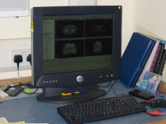
In the latter half of the eighties these solutions began to crystallise. Computers were being introduced across the NHS and their impact was not lost in radiotherapy. Pads of tracing paper were replaced with the first generation of planning computers. The simple “Bentley-Milan” algorithms could account for patient outlines accurately and speedily and optimising different beam configurations became practical. Consideration of Organs at Risk, as defined by the various International Commission on Radiation Units (ICRU) publications, became increasingly relevant. Recognition of the importance of delineating the target volumes and protecting normal tissue required improved imaging and this was provided by the new generation of CT scanners. In the nineties these were shared facilities with diagnostic radiology departments. However, the improvements provided by this imaging, enabling accurate 3-dimensional mapping of the disease with adjacent normal tissues and organs at risk, dictated their inclusion into every radiotherapy department soon after the millennium. The added bonus of using the grey scale pixel information, or Hounsfield numbers, to calculate accurate radiation transport distributions soon followed when the mathematical and computer technology caught up with the task. The value of MR and Positron Emission Tomography (PET) imaging was also recognised in the diagnosis, staging and planning of radiotherapy and the new century saw all of these new technologies embedded within the department.

Mould room technology was also improving with “instant” thermoplastic immobilisation shells replacing the uncomfortable plaster and vacuum forming methods. Custom shielding with low melting high density alloys was becoming routine and it was not long before these techniques were married with the emerging CT planning to provide “conformal” treatments.

picture 067LinAc technology also received added impetus. Computers were firstly coupled as a front end to conventional LinAcs as a safety interface to reduce the potential for “pilot error”. Their values were soon recognised by the manufacturers and were increasingly integrated into the machine, monitoring performance digitally and driving the new developments of Multi Leaf Collimators (MLC) and On Board Imaging (OBI).

The dominos for the radiotherapy renaissance were stacked up, but it needed the radiographers, clinicians and scientists to decide on the direction of travel. Computer power coupled with advanced electro-mechanical design had transformed MLC efficiency and resolution. Conventional conformal planning was now progressively superseded by sophisticated planning algorithms using merged CT and MR images. Intensity Modulated RadioTherapy (IMRT) had arrived in its evolving guises of multiple fixed field, dynamic arc therapy (RapidArc) or Tomotherapy. Whichever technique, they all offered the radiotherapy “Holy Grail” of providing three dimensional homogeneous dose distributions conformed to the Planning Target Volume (PTV) whilst achieving the required dose constraints for organs at risk and normal tissue preservation.

The tools had arrived, but an infrastructure to introduce these “toys” safely into a complex clinical background had also developed alongside. Quality standards (ISO9000), Clinical Trials, Multi Disciplinary Teams and Peer Review were governance mandates for all oncology departments and radiotherapy was leading the way. In forty years, radiotherapy had lost the “Cinderella” image and had been invited back to the clinical ball. Noticeably, breast and prostate adenocarcinoma constituted half of the radical workload.

The question remains of how and why did this transformation occur? Obviously the developing computer power and technology were the pre-requisites for many of the developments, but a key catalyst was the foresight of all of the radiotherapy family from which enduring friendships have been forged. The working lives of the clinicians and physicists involved in radiotherapy planning have probably changed more dramatically than those of any other medical and paramedical groups over the last 35 years.

We may have retired, but we still cogitate about the future direction and science behind this developing and essential cancer treatment and look forward to our younger colleagues enjoying their careers as much as we enjoyed ours.


About David Morgan

david morganDr David A L Morgan began training in Radiotherapy & Oncology as a Registrar in 1977, and in 1982 was appointed a Consultant in the specialty in Nottingham, continuing to work there until his retirement in 2011. He joined the BIR in 1980 and at times served as Chair of its Oncology Committee and a Member of Council. He was elected Fellow of the BIR in 2007. He is author or co-author of over 100 peer-reviewed papers on various aspects of Oncology and Radiobiology.


About Andrew Moloney

andy moloneyAndy Moloney completed his degree in Physics at Nottingham University in 1980 before joining the Medical Physics department at the Queens Medical Centre in the same city. After one year’s basic training in evoked potentials and nuclear medicine, he moved to the General Hospital in Nottingham to pursue a career in Radiotherapy Physics and achieved qualification in 1985. Subsequently, Andy moved to the new radiotherapy department at the City Hospital, Nottingham, where he progressed up the career ladder until his promotion as the new head of Radiotherapy Physics at the North Staffordshire Royal Infirmary in Stoke-on-Trent. Over the next twenty years Andy has acted as Clinical Director for the oncology department and served on the Radiation Physics and Oncology Committees at the BIR and was appointed a Fellow in 2007. He has been the author and co-author of multiple peer reviewed articles over the years prior to his retirement in 2017.


When medical physicists wore white coats

NHS

Dr Edwin Aird shares his memories of the revolution he has experienced working in the medical physics department.


Edwin Aird portrait

While I was an undergraduate at Newcastle University (1962), the 2nd year honours group was invited to visit the Medical Physics Department at Newcastle General Hospital. I was so impressed with the department and the range of things they were doing and the application of physics to medicine that I wrote to Professor Frank Farmer (Head of Department at that time) to ask about working there. He responded with a proposal that I apply for a special research grant that he was hoping for locally and that I write again after my graduation.

The following year I got in touch with Frank Farmer again and was accepted on an MSc grant to study “In-homogeneities in Radiotherapy”; one year in the first instance (on a grant of about £700).

Meeting 1970 1

I can’t now remember my first day exactly; not sure what office I had, but I do remember the Professor’s insistence on donning a white coat (provided and laundered by the hospital) when arriving at work, This ‘uniform’ was thought to be a vital sign to patients that physicists were part of the ‘clinical’ team.

I divided my time in that first year between research and clinical work (interestingly, this was part of the philosophy of some of the early physicists, many of whom in the 1930s and 40s had transferred from academic physics, including Frank Farmer); the situation now couldn’t be more different.

Fig 1

Image 1 The gantry mounted linear accelerator at Newcastle General Hospital [ref 2]

So, on the routine side, I began to learn about radiotherapy: its planning and treatment. Newcastle at that time had two cobalt machines (Mobaltron-60s) and two linear accelerators as well as two Marconi 250kV sets and a Philips SXT (Superficial X-rays). This latter set I used extensively a little later in my career to optimise the characteristics of the Farmer chamber (image 1).

The main linear accelerator was manufactured by Mullard (a branch of Philips), which Frank Farmer had installed as the first gantry-mounted 4MV linac (image 2). There were a few stories about the installation of this linac. In particular I remember this: the team had reached the point where they needed to produce X-rays so needed a high atomic number transmission target. Someone found a sovereign, but forgot the amount of heat produced when the high energy electrons hit it. The sovereign promptly melted and fell out of the linac head. (This linac went on to perform 25 years’ service).

Fig 2

Fig 2 Newcastle Simulator (Frank Farmer at Controls) [ref 3]

For those physicists who find quality assurance (QA) on linear accelerators today a huge burden, my recollection of the checks we performed on this linac amounted to: dose measurements and field size checks. There was little attempt to measure flatness since the accelerated electron beam wasn’t bent before striking the target, so no perturbations in intensity across the field were expected.


Fig 3

Fig 3 A Head and Neck Xeroradiograph showing potential. Treatment volume [ref 3]


Treatment planning was done by hand, often mainly using % depth dose, but also with some isodose curves for more complex plans on tracing paper; on to an outline of the patient performed using lead wire and other devices, e.g. callipers. Newcastle was unique in having developed a home-made simulator (using a radiotherapy SXT unit mounted on a gantry (image 2) that allowed an image to be viewed on a Xerox plate immediately without the need for film development (ref 3, see also image 3).

The first computer in the department, which was to revolutionise treatment planning, came to Newcastle in the early 1970s and was called a PDP 8, the ‘Rad8 system’ developed by Bentley and Milan. (image 4 [ref 4]).

Radium tubes and needles were still extensively used at this time for intracavitary and interstitial brachytherapy. I was also required to calculate the dwell times for gynae radium insertions (using ovoids and central vaginal sources with a modified Manchester system for the gynae and radium needles, mainly in back of tongue with radiographs and Paterson Parker tables). Following the calculation of the radium dwell times I would then go up to the ward to discuss the removal times, for each patient, with sister on the ward. Amazing now to think how much radioactivity (up to 105mg – equivalent to approximately 389 MBq-or more) for gynae insertions was handled by several different groups of staff at that time.

In those early years I also learnt the elements of radiation protection and nuclear medicine. Radiation Protection (guided by a ‘Code of Practice’; an excellent document that was used to help write the ‘Guidance Note’ now used under Ionising radiation Legislation) was regionally organised. Very early in my career (there were only two of us to perform the external radiation work: myself and MJ Day), I found myself organising visits within Northumberland, Cumbria and parts of Durham. (See The Regional Centre below). These were more as inspections than to make many measurements (not the very extensive QA that is performed now, although we did use film or image intensification – only recently developed – to look for faults in lead aprons; and we measured exposure levels in and out of beam with the ‘37D’ (Pitman) dosemeter (originally developed by Sidney Osborne as an excellent versatile instrument for exposure measurements ref 5). I learnt a pattern of inspection of barriers, filtration, lead aprons etc. very quickly. [In the early 1970s the NRPB decided to do their own inspections and were surprised to find that, because the HPA had organised things so well there was little need for NRPB to get too heavily involved].

My memories of nuclear medicine: incredibly slow rectilinear scanners, prior to the commercial development of gamma cameras; kidney function (renogram) using two ‘D’-shaped detectors (scintillation) with manual optimisation of their positions connected to count rate meters and chart recorders.

The regional centre

I’m not sure how many hospitals this covered in my early days in Northumberland, Tyneside, Durham and Cumbria. I know when Keith Boddy implemented Frank Farmer’s plan to have a small physics department (where gamma cameras were installed in district general hospitals) there were thirteen hospitals with physics departments in the region. Prior to this – from about my 3rd year at Newcastle – I was delegated to look after the small centre in Carlisle where there were two Marconi 250kV sets, an SXT and some iodine treatment. In the 1950s this had been the job of Jack Fowler who used some of his time to study arc therapy on 250kV (see ref 6).

Rad 2014 photo

Edwin Aird (right) receiving the BIR Sylvanus Thompson Award from Andy Beavis

In those early years I also found time to help develop differential X-ray absorptiometry to measure antimony in the local Tyneside Antimony workers’ lungs (organised by the Newcastle University Department of Industrial Health). I was also able to build on this experience to develop my own equipment, using characteristic X-rays, to measure bone mineral in the femur. This clinical work allowed me to meet a new set of clinicians (other than radiotherapists – as Clinical Oncologists were then called – and radiologists) involved with bone loss in patients: kidney specialists, endocrinologists and geriatricians. Commercial equipment has since been developed to perform bone mineral and body composition measurements (GE Lunar, Hologic, and Norland).


References:

  1. Aird EGA and Farmer FT. The design of a thimble chamber for the Farmer dosemeter. Phys Med Biol 1972 17: 169–174. https://doi.org/10.1088/0031-9155/17/2/001
  2. Day MJ and Farmer FT. The 4 MeV Linear Accelerator at Newcastle upon Tyne. Br J Radiol 1958; 31: 669–682. https://doi.org/10.1259/0007-1285-31-372-669
  3. Farmer FT, Fowler JF and Haggith JW. Megavoltage Treatment Planning and the Use of Xeroradiography. Br J Radiol 1963; 36: 426–435. https://doi.org/10.1259/0007-1285-36-426-426
  4. Bentley RE and Milan J. An interactive digital computer system for radiotherapy treatment planning. Br J Radiol 1971; 44: 826–833. https://doi.org/10.1259/0007-1285-44-527-826
  5. Osborn SB and Borrows RG. An Ionization Chamber for Diagnostic X-Radiation. Med.Biol1958; 3: 37–43. https://doi.org/10.1088/0031-9155/3/1/305
  6. Fowler JF and Farmer FT. Measured Dose Distributions in Arc and Rotation Therapy: A Critical Comparison of Moving and Fixed Field Techniques. Br J Radiol 1957; 30: 653–659. https://doi.org/10.1259/0007-1285-30-360-653

About Dr Edwin Aird

Edwin Aird was Head of Physics at Mount Vernon Hospital from 1988-2012 and Head of Radiotherapy Physics at St Bartholomew’s Hospital from 1985-1988. He was Head of Radiotherapy Physics at Newcastle General Hospital from 1980-1985. He is an IPEM Chief Examiner 1994-1997 (Physics Training Scheme), FRCR (RT) Examiner 1989-1993, Radiotherapy Degree (External Examiner (Liverpool University): 1993-1996. He is a IAEA: Qualified Expert: 1999-2005 and an LH Gray Trustee: 1999-2003.

He was awarded the BIR Roentgen Prize in 2005 and delivered the Silvanus Thompson: Award and Eponymous Lecture 2013.

The case of the missing fingers!

NHS

Professor Roger Dale remembers how he got his first job in medical physics and how he thought he’d discovered a radiation martyr.


Roger Dale circa 1966

Anxiously seeking a job in medical physics on completion of my first degree in 1966 I quickly became aware that basic grade physicist positions in large centres were difficult to find and, for a while, I was unsure what to do. Being out of work I wrote in some desperation to a (very small) radiotherapy centre in Kent pointing out my predicament and asking if I could join as a porter until such time as I could obtain a physicist position in a larger department. To my great surprise I received a phone call a day or two later from the head radiotherapist (Dr B) inviting me along for an informal chat with him, during which it transpired that the hospital had no requirement for any more porters but did have a vacant establishment for a radiotherapy physicist at principal grade! The principal post had already been offered to a gentleman in New Zealand but it would take a month or two before he could take up the position. Therefore, as there was no physicist in post at that time, Dr B suggested that I join as an acting-temporary(!) basic grade until the principal appointee arrived in the UK. Needless to say, I agreed without hesitation.

The necessary paperwork was sorted out remarkably quickly (the old personnel departments always seemed notably more efficient than the burgeoning HR empires which later followed) and my career in medical physics began, albeit rather shakily. My only ‘supervision’ came from occasional conversations with Mr W, the Chief Technician, whose own duties were entirely focused on running the film badge and thyroid uptake services. He was not at all involved on the radiotherapy side of things so I spent many hours buried deep in the standard radiotherapy physics textbooks of the time. That reading reinforced my desire to stay in medical physics because here were the seemingly abstract physical and mathematical concepts encountered during my degree studies being successfully applied to highly relevant clinical issues. Amongst other things I brushed up on the fundamentals of radium dosimetry, this being necessary since Dr B performed several radium implants each week (remote afterloading systems were only just being introduced back then) and, as I was now the sole medical physicist (of sorts) within a 50 mile radius, he required me to be present during the procedures.

Dr B’s theatre sessions were an eye-opener. Apart from a certain squeamishness at witnessing surgery for the first time, I found his implantation technique quite scary since, although a full range of surgical implements and manipulators were at his disposal, he had a habit of giving all the radium needles a push with his fingers. Worse, it was impossible not to notice that several of his fingers were in fact missing! Even a greenhorn like me knew that physically touching radioactive sources was definitely a practice not to be recommended and the fledgling scientist in me began to ponder on cause and effect.

For several days it worried me that Dr B might be paying a very high price in order to pursue his noble vocation and I was unsure how (or if) I should air my concerns, especially as my status as an unsupervised acting-temporary basic grade physicist of just a few weeks’ standing hardly conferred much authority. Eventually I plucked up the courage to speak to the Chief Technician, telling him how convinced I was that Dr B was suffering radiation damage as a direct result of his operating technique. Mr W’s reaction was not quite what I expected. After some snorts of derision at my expense he then took some delight in pointing out that Dr B had been in the RAMC during the war. He had landed on the Normandy beaches where his jeep had hit a mine, and that was how he had lost several of his fingers. Somewhat chastened, I went away to reflect on the fact that my powers of deductive reasoning might be in need of substantial refinement.

Shortly after this awkward conversation the newly-appointed principal physicist arrived from New Zealand and, contrary to all my expectations, Dr B suggested that I stay on for a while longer to gain some first-hand experience working with the new man. This was to be a tremendous bonus as the knowledge and advice I picked up in the weeks following gave me enough of an advantage to successfully apply for a substantive post (i.e. neither acting nor temporary) in a large London centre, after which I never looked back.

Roger Dale recentToday’s NHS is nothing like the one I joined in 1966 and specialised scientist training is much more formalised and incalculably better. No one these days could be appointed in the manner that I had been but Dr B, like most other NHS professionals then and now, was motivated by good intentions and his thoughtfulness over fifty years ago put me on the path to a rich and fulfilling career in medical physics and radiobiology. I discovered later in life that Dr B had told one of his colleagues that he had helped me because he “wanted to give the lad a chance”. What he gave me was a chance that was truly exceptional and this lad has been immensely grateful ever since.


About Professor Roger Dale

Roger Dale retired from his post at Imperial College Healthcare in 2010 following an NHS career spanning 43 years. His main scientific interest has been the development of radiobiological models which can be used to quantitatively assess the biological impact of radiotherapy and other cancer treatment modalities. He is widely published and the clinical significance of his work has been recognised through the award of a number of prestigious scientific prizes and through his  parallel appointment, in 2005, as Professor of Cancer Radiobiology in the Faculty of Medicine at Imperial College. He continues to be involved in research and teaching.

My first day in radiotherapy physics: reflections of a medical physicist

NHSIn 2010 Karen Goldstone was awarded the MBE for her services to healthcare. Here she reflects on the primitive tools used for radiotherapy patient outlines back in the 1970s and remembers the wise advice she was given on her first day as a radiotherapy physicist.

BIR

I started work in the NHS as a Hospital Physicist in 1970. Prior to that I did the MSc in radiation Physics based at Middlesex Hospital. When doing a placement in nuclear medicine, computer tapes had to be taken to University College about a fifteen minute walk to the other side of Tottenham Court Road and fetched the next day hopefully having run successfully.

In my first post I expected to be doing mainly diagnostic radiology physics but discovered that that was rather a luxury field and so most of my time was spent doing radiotherapy physics. Those were the days when patient outlines were taken using a strip of lead or a flexicurve and planning was done using tracing paper and coloured pencils or biros. There was no computer planning of course and we only had one calculator with a paper roll print out so slide rules were in constant use. The main piece of advice I remember receiving on my first day was that if I discovered I had made a mistake I should own up to it straight away and not seek to cover it up – very wise words.

When not doing radiotherapy physics many hours were spent reading out film densities produced using our homemade “Ardran Cassette” in order to check kVp. This was the beginning of setting up a quality control programme for X-ray units. Another time-consuming activity was sealing lithium borate powder into plastic capsules in order to measure dose to radiologists, carrying out various procedures under fluoroscopic control, and subsequently reading the doses received.

Although diagnostic radiology physics was not seen as important it was an exciting time and I was fortunate enough to hear Godfrey Hounsfield give the 1972 MacRobert Award lecture on “Computerised Transverse Axial Tomography” – an invention that has revolutionised diagnostic imaging.

I started my second post in 1974 in a smaller department but with responsibilities in other, far-flung, hospitals. Here I was the radiotherapy physicist (the only one) and also covered diagnostic radiology and radiation protection, but because it was a smaller department and staff had to be versatile I also did some nuclear medicine and even once some ultrasound.

In the peripheral hospitals in my patch one was still using wet developing, one using just a fluorescent screen for fluoroscopic procedures and one an image intensifier viewed not via a camera but via a mirror arrangement.

How times have changed!


About Karen Goldstone MBE

I worked for forty years in the NHS, in radiotherapy physics, diagnostic X-ray physics and all aspects of radiation protection. In 1983 I set up the East Anglian Regional Radiation Protection Service (EARRPS) based at Addenbrooke’s Hospital in Cambridge, and ran it for almost 30 years. I was both a Radiation Protection Adviser and Laser Protection Adviser. I gave physics lectures to radiologists and was a physics examiner for FRCR both in the UK and Malaysia. With colleagues in EARRPS and Cambridge University I ran a number of Radiation Protection Supervisor courses and gave countless IRMER courses to reluctant clinical staff. I was exceedingly surprised to be awarded the MBE in 2010 for services to healthcare.

Since retiring I have taken up rowing and become a Level 2 rowing coach; I have given two courses on Radiation and Health to the University of the Third Age in Cambridge and am otherwise kept busy with my garden, allotment, grandchildren and church activities. I am still involved on one or two committees for medical physics and radiological protection.

Hats off to Sir Peter Mansfield (1933-2017)

13-sir-peter-mansfield-2003

Sir Peter Mansfield left school with no qualifications to become one of the most eminent scientists in the world of physics. Here, Dr Adrian Thomas pays tribute to the man who lived through World War Two and with dogged determination forged his way in science to become a distinguished and recognised physicist who played a major part in the story of MRI.


Sir Peter Mansfield was born on 9 October 1933 in Lambeth in London, and grew up in Camberwell. His mother had worked as a waitress in a Lyons Corner House in the West End of London, and his father first worked as a labourer in the South Metropolitan Gas Company, and then as a gas fitter. Mansfield recounted being sent with other children on a holiday to Kent for disadvantaged London children by the Children’s Country Holiday Fund.

Peter Mansfield was 5 years old when the war broke out in 1939. He remembers standing with his father at the entrance of an air raid shelter watching anti-aircraft shells exploding around German bombers caught in the searchlights. As the Blitz intensified he was evacuated from the dangers of the capital, as were so many other London children. With his brother he was sent to Devon, where he was assigned to Florence and Cecil Rowland who lived in Babbacombe, Torquay. The Rowlands were called Auntie and Uncle, and Mansfield  attended the nearby junior school. Cecil Rowland was a carpenter and joiner by trade, and encouraged Peter to develop his practical skills by giving him a toolbox, and tools were slowly acquired. He obviously obtained some proficiency since with some guidance he made several wooden toys which he was able to sell at an undercover market and a toyshop in Torquay. His life was not without danger even outside London, and in early 1944,whilst out playing, he saw a German twin-engined Fokke-Wulf plane flying at rooftop level. The tail gunner was spraying bullets everywhere, and he rapidly took shelter behind a dry-stone wall.

On his return to London his secondary schooling was at Peckham Central, moving  to the William Penn School in Peckham. Shortly before he left school at 15 he had an interview with a careers adviser. Peter said that he was interested in science, and the adviser responded that since he was unqualified that he should try something less ambitious. He was interested in printing and so took up an apprentice in the Bookbinding Department of Ede and Fisher in Fenchurch Street in the City of London, and whilst there he took evening classes.   Developing an interest in rockets he was offered a position at the Rocket Propulsion Department (RPD) at Westcott, near Aylesbury.

In 1952 he was called up into the Army for his National Service, where he joined the Engineers. The Army allowed him to develop his interest in science. On demobilization he returned to Westcott and completed his A levels. This enabled him to apply for a special honors degree course in physics at Queen Mary College in London. In 1959 he obtained his BSc, and three years later he was awarded his PhD in physics. From 1962 to 1964 he was Research Associate at the Department of Physics at the University of Illinois, and in 1964 was appointed Lecturer at the Department of Physics at the University of Nottingham.

During a sabbatical in Heidelberg in 1972 Mansfield corresponded with his student, Peter Grannell in Nottingham, and became interested in what became MRI, presenting his first paper in 1973 at the First Specialized Colloque Ampère. Mansfield developed a line scanning technique, and this was used to scan the finger of one of one of his early research students, Dr Andrew Maudsley. The scan times required for these finger images varied between 15 and 23 minutes. These were the first images of a live human subject and they were presented to the Medical Research Council, which in 1976 was reviewing the work of various groups including those in Nottingham and Aberdeen.

13-terry-baines-peter-mansfield-and-andrew-maudsley-c1974

In 1977 the team at Nottingham, which included the late Brian Worthington, successfully  produced an image of a wrist. The following year Mansfield presented his first  abdominal image. In 1979 Peter Mansfield was appointed Professor of Physics at the University of Nottingham. As the Nobel Committee emphasized, the importance of the work of Peter Mansfield was that he further developed the utilization of gradients in the magnetic field. Mansfield demonstrated how the signals could be mathematically analyzed, which resulted in the development of  a practical  imaging technique. Mansfield also demonstrated how to achieve extremely fast imaging times by developing echo-planar imaging. This is all very impressive for a boy who left school at 15 with no qualifications.

13-sharing-an-amusing-tale-with-paul-lauterbur-2003

Peter Mansfield was awarded many prizes and awards including:

the Gold Medal of the Society of Magnetic Resonance in Medicine (1983); Fellow of the Royal Society (1987); the Silvanus Thompson Medal of the British Institute of Radiology (1988); the International Society of Magnetic Resonance (ISMAR) prize (jointly with Paul Lauterbur)(1992);  Knighthood (1993); Honorary Fellow of the Royal College of Radiology and Honorary Member of the British Institute of Radiology (1993);  the Gold Medal of the European Congress of Radiology and the European Association of Radiology (1995);  Honorary Fellow of the Institute of Physics (1997); the Nobel Prize for Medicine together with Paul Lauterbur (2003);   Lifetime Achievement Award presented by Prime Minister Gordon Brown (2009).

His autobiography The Long Road to Stockholm, The Story of MRI was published in 2013. This is an interesting read, particularly in relation to his early years, and is recommended reading for everyone interested in the radiological sciences. This is a revealing account of a remarkable life. Whilst we may discuss the complexities of the development of MRI and exactly who should have received the Nobel Prize, there can be no doubt about his major contributions. MRI has made, and is making major contributions to health care. He died age 83 on 8 February 2017.

The University of Nottingham has set up an online book of condolence http://www.nottingham.ac.uk/news/sir-peter-mansfield/

About Dr Adrian Thomas, Honorary Historian BIR

Dr Thomas was a medical student at University College, London. He was taught medical history by Edwin Clarke, Bill Bynum and Jonathan Miller. In the mid-1980s he was a founding member of what is now the British Society for the History of Radiology. In 1995 he organised the radiology history exhibition for the Röntgen Centenary Congress and edited his first book on radiology history.

He has published extensively on radiology history and has actively promoted radiology history throughout his career. He is currently the Chairman of the International Society for the History of Radiology.

Dr Thomas believes it is important that radiology is represented in the wider medical history community and to that end lectures on radiology history in the Diploma of the History of Medicine of the Society Apothecaries (DHMSA). He is the immediate past-president of the British Society for the History of Medicine, and the UK national representative to the International Society for the History of Medicine.

See more on the history of radiology at http://www.bshr.org.uk


Top tips for honest science messages in the media

13-kate-elliottScience is often misrepresented in the media. The BIR supports the charity Sense about Science in their call for all research to be openly and honestly reported. This year we supported one of their Voice of Young Science workshops called “Standing up for Science” held on 16 September 2016 in London.

Here, Kate Elliott, Medical Physicist at  Mount Vernon Cancer Centre was one of three lucky BIR members to attend the workshop which gave young researchers top tips and advice on how to get their scientific messages across as clearly and accurately as possible.


I hate speaking in public and even the thought of writing this article terrified me. Why then, you might ask, did I apply to go on the Standing up for Science media workshop?

I often get annoyed at the coverage of science in the media and the misuse of statistics and results. Recently, the Brexit “debate” has left me ranting at friends, and I often find myself defending junior doctors on social media. When I received the email from BIR advertising the media workshop, it struck me as an opportunity to learn what I could do to positively influence the public perception of science, and to hear first-hand from journalists about their involvement.

The first session consisted of a panel of three scientists who told us of personal experiences with the press and offered advice based on this. An example which stood out to me as a healthcare scientist was Professor Stephen Keevil’s use of the media to highlight a problem with a new EU directive on physical agents[1], which could  have caused problems for MRI. Politicians took heed of his criticism, and effected a change to the directive in Brussels. This was a great example of how the media can be used effectively to influence policy – something that is likely to become increasingly important in the next few years.

The second session was a panel of three journalists, who explained their daily process for13-standing-up-for-science-workshop-sept-2016selecting and pitching stories. Science stories are selected based on interest, accessibility, and importance. These are pitched to the editors, who decide which ones to take further. The journalists pointed out that their duty is to their audience, not to science. Unfortunately, science has to compete with news on David Beckham’s haircut. Time constraints are also a problem. They write multiple articles a day (I’m three weeks and counting on this one…), so it’s important for scientists to be available to discuss their research on the day it’s published.

The third panel was about the nuts and bolts of how to interact with the media, and recommended campaigns such as Sense about Science’s “Ask for Evidence” campaign.

I left the event with the following advice to keep in mind:

  • If you disagree with something: speak out. If the public only hears one side of the story, that’s the side they’ll believe.
  • Stick to a few key points. Get those across, even if it means having to ignore questions or turn them around in an infuriatingly politician-like way!
  • Be available. If you’ve put out a press release, you need to be able to respond quickly. Journalists work to very stringent time scales, so being available in a week’s time is going to be too late.
  • Talk to the public. Attend events such as Pint of Science, or become a STEM ambassador, because that will really help you learn to speak in layman’s terms and get you used to answering obscure questions.
  • Get training. If not full media training, a workshop like this is a really good way to be slightly more prepared – and you get to hear about all the interesting science other people are involved in!

Image: BIR members  Jim Zhong, Kate Elliott and Maureen Obioha Agwanihu who attended the workshop

[1] https://www.myesr.org/html/img/pool/MRI-Report-Stephen-Keevil.pdf