Radiography, Research, and You

Kim Mason head and shoulders image
Kim Mason

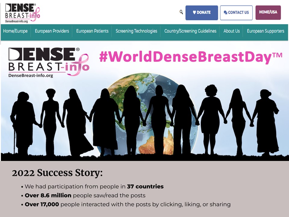
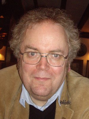
Kim Mason, an Audit and Research Radiographer for Mid Yorkshire Teaching Hospitals Trust, talks about their role as well as the value of radiographer engagement in research activities and how to get involved.

In December, 1895, Wilhelm Röntgen would x-ray the hand of Anna Bertha Ludwig, his wife, using a photographic plate. The new discovery lit a fire in the scientific community, and was so sensational that in the following year over 1,000 articles would be published on the topic of X-rays. Over the next 130 years, medical imaging has undergone many varied evolutions to become a cornerstone of modern-day medicine. All beginning with that first piece of research.

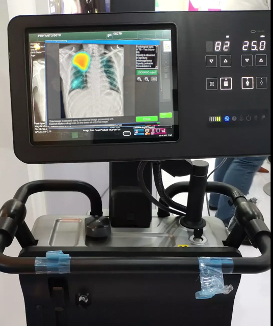
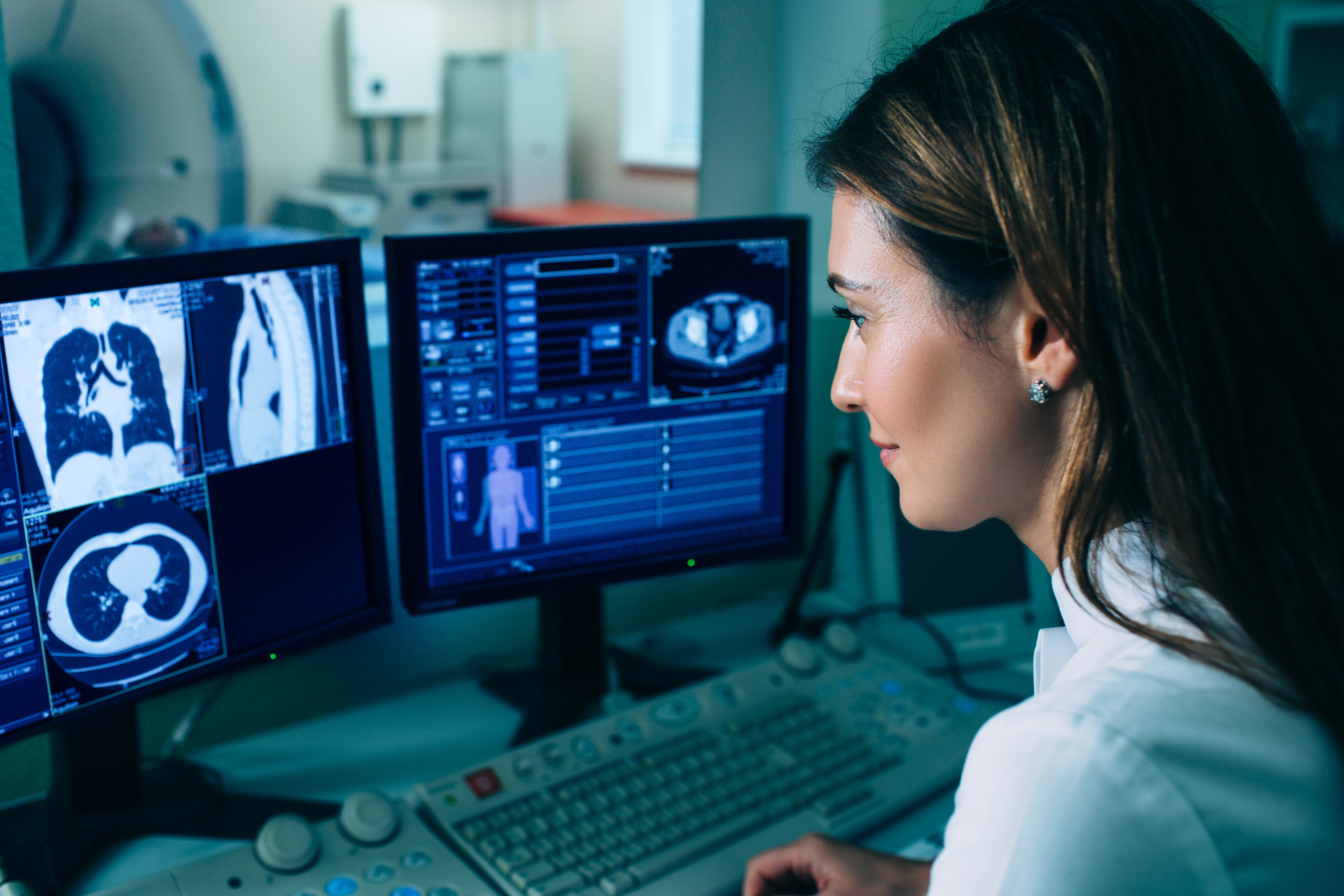
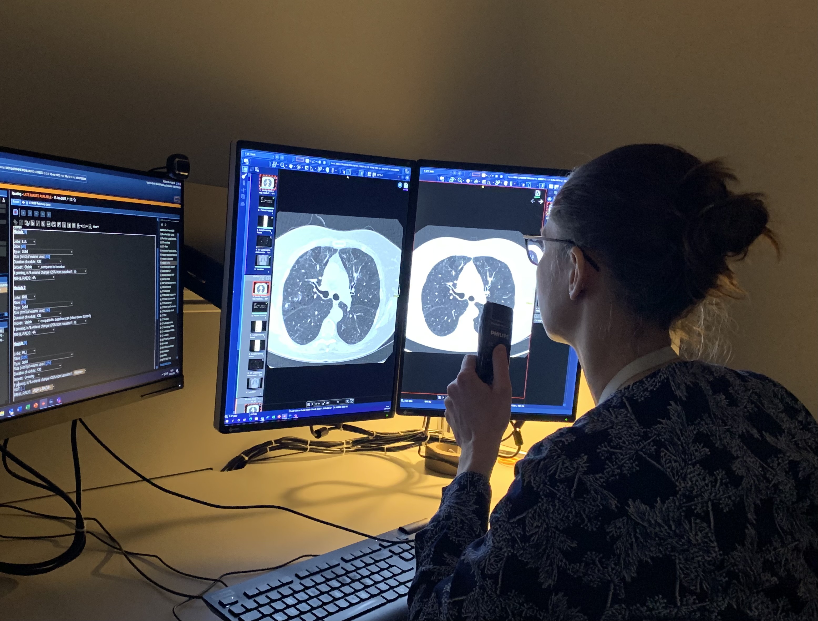
Radiography has exploded into a variety of modalities and specialisms from CT to Ultrasound to MRI; all driven by research and development. That is where I come in.

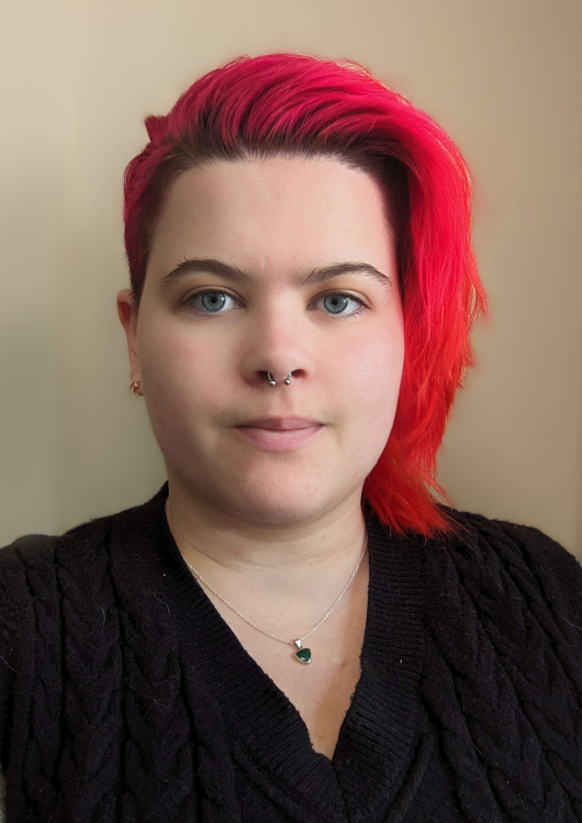
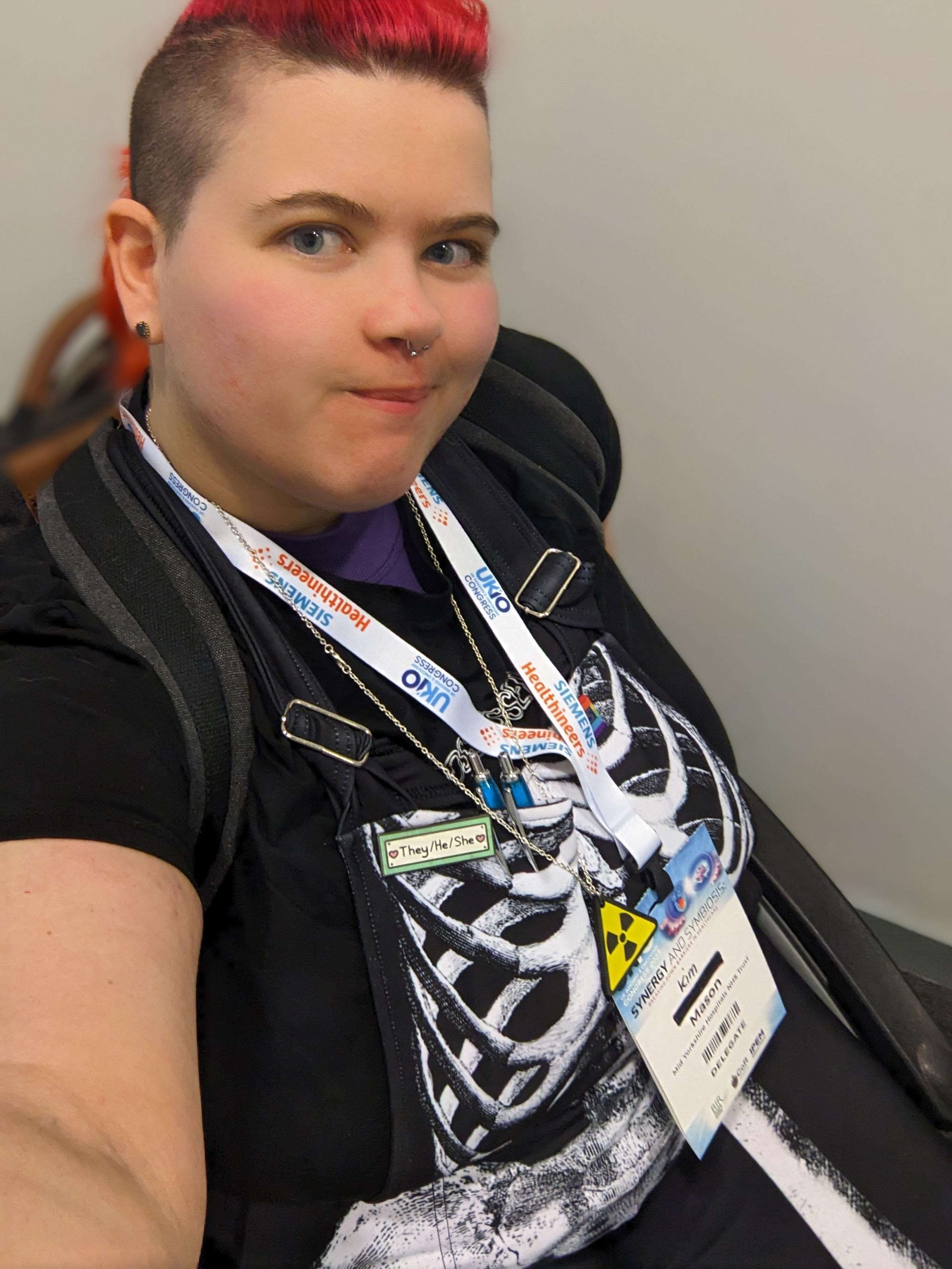
Hi, I’m Kim and I am an alternative-styled, funky-haired, septum-pierced, disabled Audit and Research Radiographer. That is to say, I’m fairly easy to pick out of a line up. I’m also passionate about education, research, and (of course) radiography. This, I’ve been told, is also very easy to pick up on.

I graduated with my radiography degree in 2018 and since then I’ve worked in a wide variety of departments, from plain film to nuclear medicine. Since I found something to love in every modality it never really mattered which one I was in. Prior to my current position, I’ve spent time as a Radiation Protection Supervisor, and well as a trainer for graduate and post graduate radiographers. Now I’m in Audit and Research, which is far different to anything I’ve done before.

So, what is an Audit and Research Radiographer?

Kim sat in their wheelchair at the UKIO 2023 conference

My position is entirely based in research so I do not currently undertake any non-research imaging. However, I’m not entirely non-clinical. I get trained to undertake research scans on new equipment as required for me to carry out the necessary imaging for research trials. I also provide support to all trials requiring access to our radiography department, regardless of the imaging modality.

My job requires that I read a lot of trial protocols so that I can determine whether we have the necessary resources to undertake the trial. It can be hard work, and for me to be effective I have to have a good understanding of how each of our trusts’ imaging modalities function. I also need to have strong communication skills, especially in regards to explaining radiography perspectives to multi-disciplinary team members who may have little to no understanding of imaging. More than this, I need to be able to form and maintain good relations with all sides to ensure that effective communication can take place.

The work I do allows for better outcomes between radiology and research departments. It also affords me the opportunity to be a part of progress, seeking out better practice or improved technology for the future of imaging. In addition, I am able to undertake my own research, working towards my goals of further academic qualifications.

My role is important. I feel that I and my work contributions are valued by both the radiology and research teams. My pathway for personal development is clear and I am able to see the benefits in the set-up and management of the trials my Trust undertakes.

What is Radiography research and why is it important?

There is often research aimed at improving and advancing the field of diagnostic imaging. Currently there are trials into new scanning techniques; new equipment with the potentials for dose reduction and/or improved image quality and patient experiences; and the use of AI in imaging and reporting.

The benefits of improvements in our field are numerous. Every radiographer wants to give patients the best experience they can, however this is often at odds with the nature of our job. Requiring patients to hold uncomfortable positions when they are in pain or worried; perhaps having to go through narrow tubes which are sometimes incredibly loud; needing injections which makes them feel weird or mean they are radioactive and have to keep distance from other people at a time when they could really do with support. With research, we can aim to improve these experiences; reduce scan times or radiation exposures, wider bore scanners or open scanners, even finding new imaging or testing which removes the need for the ionising radiation all together!

Patients are at the heart of the NHS, and diagnostic imaging is often an area in which a good patient experience can be harder to provide (that is not to say that we don’t try!). With research, we have the potential to make those improvements to service, to provide for our patients in the way which we want to and the way that they deserve.

How can I get involved?

Often, radiography research is overseen by radiologists, doctors and orthopaedic surgeons, but there is no reason why radiographers shouldn’t also get involved. As we are the ones who use the equipment on a daily basis, consent and care for the patients during imaging, and come into contact with the faults and issues. Our profession contains a wealth of knowledge which can be used to improve all aspects of radiography.

You don’t need any academic qualifications to get started with research activities. In fact, many radiographers image research patients without being privy to the research aims. In a busy department, such patients are treated the same as any other in most regards. If you do image a research patient, perhaps look into the trial itself. As well as being interesting additional information, it can be used as material for CPD in the form of a case study or reflective piece. You may also discover potential ways to improve the patient experience within your department and help to enact future change.

Look into what your hospital requires for research involvement. The Good Clinical Practice (GCP) qualification, which is usually necessary, can be found for free as an online e-learning module. The NIHR website provides a lot of helpful information for getting started. NIHR also provides help for those who are wanting to gain further academic qualifications, such as through grant applications for fellowship awards. These are highly competitive but allow better access for NHS employees to undertake Masters or Doctorate level qualifications. The NIHR also run conferences for those who are new to research but interested in how they can take part.

You could also look at taking part in your department’s audits. Audits are a great way to check in on the health of your department, what you are doing well, and what you can improve on. Audit skills can also overlap with those necessary for research work, as well as provide possible avenues for research within your department.

How do I get a research job?

The roles of radiographers in research are expanding. Some hospitals offer clinical research radiographer positions, which give additional responsibility to train for and undertake specialised research imaging, often alongside a multi-disciplinary team. Other trusts may offer training for those wanting to aid research trials.

For research-specific roles, take a look at NHS jobs. You will find posts for Research Radiographers or Research Clinical Practitioners/AHPs. When I applied for my Audit and Research radiographer post, I had no specific research skills however I was well versed in audits and had learned about the processes of research in my own time. Enthusiasm goes a long way when applying for research roles, we need radiographers who are driven and raring to get stuck in.

There is so much experience and knowledge that radiographers have to offer research, and there’s so much improvement and advancement to be received in turn. I strongly encourage any radiographers to give it a try. You never know, you may get hooked!

To submit your research to a BIR journal find out more here:

BJR https://www.editorialmanager.com/bjr/default2.aspx

BJR Case|reports https://www.editorialmanager.com/bjrcr/default2.aspx

BJR|Open            https://www.editorialmanager.com/bjro/default2.aspx

About Kim Mason

Kim Mason is a HCPC registered diagnostic radiographer, graduating from the University of Leeds with a 1st Class BSc Honours in Diagnostic Radiography in 2018. They have experiences in education both inside and outside of radiography, and have a passion for improving the radiography services in the UK.

Kim has multiple chronic conditions, and as such, they are an ambulatory wheelchair user. This has given them keen insight into the experiences of patients within the radiography department, having undergone imaging in most modalities as a patient. They have a vested interest in educating the public about radiography, and educating radiographers on improvements to patient care. 

Artificial Intelligence Embedded Imaging Modality

In the third blog of her series on AI and the radiographer, Shamie Kumar explores the impact on the radiographer when AI is integrated within an imaging modality.

In previous BIR blog posts, I have explored how AI is integrated into PACS with the AI outputs seen on radiology systems, and whether non-reporting radiographers could learn and benefit from AI. The question to explore in this blog is when AI is integrated within an imaging modality itself and how that may impact a radiographer.

AI embedded into a portable digital X-ray machine

Radiographic images are acquired in multiple modalities within different patient pathways. I will explore how AI embedded into a portable digital X-ray machine might change and affect how the radiographer works and learns.

Every radiographer is trained to take X-rays on portable machines and this is a core skill and it is an adapted technique compared with dedicated static X-rays rooms. It is unique in the sense patient positioning can vary depending on the environment and situation, whether this be on a ward or in A/E resus. Patient’s conscious level and mobility can vary, often supine and not all being cooperative. There can be situations where other healthcare professionals (HCP) are in proximity of the patient being imaged due to the image being acquired outside of the main radiology department.

AI output

Some hospitals have adopted digital portable X-ray machines to provide an instant image, the radiographer can see the chest X-ray immediately after exposure and decide whether the image quality is optimal. As AI becomes integrated within the modality, in this instance on a portable digital X-ray machine, the radiographer will also see the AI output and findings alongside the original X-ray. Not only does the radiographer see the AI output but other HCP that are present will also have the accessibility to view the same in the given environment. As we all know, X-rays need to be reported by radiologist or reporting radiographers, but often clinicians make clinical decision before these inpatient portable x-rays reports are finalised and available on the hospital system, especially if quick intervention is required.

When AI integration is done in such a way that radiographer need not log into PACs to view the AI output and is shown on the modality once the image is acquired, all radiographers can utilise AI to its full potential. The focus quickly shifts to: does the radiographer have the relevant education and training to understand the AI intended use, the AI outputs, what are the functions, features of the AI, how do they clinically interpret these images, how does AI work and what are the limitation of AI. All these questions become important when an AI is implemented; radiographers need to be trained how to use it, become familiar with the outputs, and educate others around them. If this is approached robustly, it will empower radiographers to learn and upskill themselves with AI being part of their daily clinical workflow, giving them the confidence to support and guide other healthcare professionals (HCPs) who also are looking at the X-ray when it acquired.

AI is an assistive tool

It’s important to recognize that AI findings are never the final diagnoses. Ultimately AI is an assistive tool, embedded within portable machines. Doctors and HCPs will also view the AI output and, with time, it will be the role of the radiographers to appropriately manage and guide other healthcare professionals.

About Shamie Kumar

Shamie Kumar is a practicing HCPC Diagnostic Radiographer; graduated from City University London with a BSc Honors in Diagnostic Radiography in 2009 and is a part of Society of Radiographers with over 12 years of clinical knowledge and skills within all aspects of radiography.

She studied further in leadership, management, and counselling with a keen interest in artificial intelligence in radiology.

An explosion in imaging: Is this the future?

Prof Simon Padley

Following the recent BIR live event Imaging explosion across the pond – causes and solutions in which the UK and USA radiology and healthcare systems were compared, DMC Radiology Reporting co-director Professor Simon Padley reflects on the UK position.

The USA often provides a window into our own future

In the application of imaging technology, we often follow trends that emerge in the US – the rise in CT and MRI utilisation are two examples, and more recently the growth of outsourcing is following a similar trajectory.

With different healthcare models, the US does not rely on central funding to replace or add to CT and MRI capacity. NHS funding comes from the government’s general revenue, and healthcare services are provided to all residents of the UK, regardless of their ability to pay. In contrast, the US medical system is a mixed system of public and private funding. It includes a combination of private health insurance, employer-based insurance, individual out-of-pocket payments, and public programs like Medicare and Medicaid, which are funded through federal and state taxes.

In 2022 there were 6.6 million CT studies on NHS funded patients according to NHS Digital, at a time when the population was reported to be 55 million. This equates to 121 studies per 1000 of the population. In the USA  this figure is more than double at 278 CT studies per 1000 of the population (84 million CT studies in a population of about 333 million CT Scans Each Year – iData Research).

And that is just CT! Official data for 2021/22 shows that between April 2021 and March 2022, NHS services in England carried out 43.8 million imaging tests across all modalities Ref. This reflects an ever-growing requirement for imaging studies to be undertaken and reported.

At the same time, the workforce needed for this activity has fallen far behind that required. We bump along the bottom of the league table for radiologists per 100,000 of the population (8.5). Europe has 13, the US 11. Couple this with complexities of pension taxation, IR35 and COVID related burnout all nudging older highly skilled and efficient radiologists towards the exit door and we have a perfect storm. Even today we have 2000 full-time consultant clinical radiologist posts unfilled across the UK. The RCR predicts a 39% workforce shortfall by 2026 (equating to 3166 full time radiologists).

This may create stress in the radiology department, but rest assured it also causes grey hair and sleepless nights for those that inhabit the carpeted management corridors. Hidden amongst every backlog of reporting there is serious pathology lying undiagnosed. When that report is provided, and the treatment options are discussed, some options will have closed, tumours will have stage shifted and outcomes will be less good. This constitutes a chief executive’s nightmare but has caught the medicolegal world’s attention. So, what are we to do?

Teleradiology and the NHS

Almost all acute trusts have turned to the services of the teleradiology community, now playing a vital role in helping to address this capacity shortfall. In the past 10 years the market has grown with a compound annual growth rate of about 10-15%.

Who are all these extra radiologists and where do they come from? Well of course, by-and-large, they are you and me. But we are a limited pool, the market rates for reporting (set by the NHS) are not great and there are only so many hours in the day.

So where can we look for additional workforce capacity and will we be allowed to access it? To allow this to happen the NHS will need to engage more readily with the solutions that are now emerging and examine the detail of how we, in the teleradiology world, are already addressing data governance and medicolegal concerns.

At DMC Radiology Reporting, we already work in partnership with many NHS Trusts. We strive to deliver fast, accurate radiology reporting with innovation and efficiency. We have a rigorous commitment to clinical governance, and we are proud of our work force of GMC-registered/FRCR-radiologists with sub-specialty interests.  Like many others, we are interested in how these problems are being addressed in the US.

About Professor Simon Padley

In 2013 Simon co-founded DMC-Radiology Reporting, which has been growing and developing ever since, focusing on sub-specialist high quality outsourced reporting.

Simon is a cardiothoracic and interventional radiologist, appointed in 1994. As a previous imaging director in the NHS for many years he developed a range of new services, most recently as lead radiologist for Royal Brompton Hospital Diagnostic Imaging Centre, opened in 2022. This facility incorporates one of the only combined interventional bronchoscopy and radiology facilities in the country.

As a Professor of Practice (Diagnostic and Interventional Radiology) since 2016, at the National Heart & Lung Institute, Imperial College London, he maintains an active academic career, publishing widely with over 220 articles in peer reviewed journals.

Can We Upskill Radiographers through Artificial Intelligence? 

Shamie Kumar describes how AI fits into a radiology clinical workflow and her perspective on how a clinical radiographer could use this to learn from and enhance their skills.

AI in radiology and workflow

We all know that AI is already here, actively being implemented and used in many trusts in seeing its real world value supporting radiology departments to solve current challenges. 

Often this is focused on benefits to radiologist, clinicians, reporting radiographers, patients, and cost savings, but what about clinical non-reporting radiographers undertaking the X-ray or scans – can AI benefit them too?

Let’s think about how AI is implemented and where are the AI outputs displayed? 

If the AI findings are seen in PACS, how many radiographers actually log into PACS after taking a scan or X-ray? Good practice is seen to have PACS open to cross-check images that have been sent from the modality. Often this doesn’t happen for various reasons but maybe it should be a part of the radiographers’ routine practice, just like post-documentation is.

Can Radiographers Up-Skill?

Given the view it does happen, radiographers will have the opportunity to look at the AI outputs and potentially take away learnings on whether the AI found something that they didn’t see initially or whether there was a very subtle finding. We all know people learn through experience, exposure, and repetition, so if the AI is consistently picking up true findings, then the radiographer can learn from it too.

But what about when AI is incorrect – could it fool a radiographer, or will it empower them to research and understand the error in more detail?

As with many things in life, nothing is 100% and this includes AI in terms of false positive and false negatives. The radiographers have the opportunity to research erroneous findings in more detail to enhance their learning, but do they actually have time to undertake additional learning and steps to interpret AI? 

CPD, self-reflection, learning through clinical practice are all key aspects of maintaining your registration, and self-motivation is often key to furthering yourself and your career. The question remains: are radiographers engaged and self-motivated to be part of the AI revolution and use it to their professional benefit with potential learnings at their fingertips? 

There have been a few recent publications that share insight on how AI is perceived by radiographers, what is their understanding, training and educational needs.

Many Universities like City University London and AI companies like Qure.ai are taking the initial steps in understanding this better and taking active efforts in filling the knowledge gap, training and understanding of AI in radiology.

Radiographers who are key part of any radiology pathway, are yet to see the real-world evidence on whether AI can upskill radiographers, but there is no doubt this will unfold with time.

About Shamie Kumar

Shamie Kumar

Shamie Kumar is a practicing HCPC Diagnostic Radiographer; graduated from City University London with a BSc Honors in Diagnostic Radiography in 2009 and is a part of Society of Radiographers with over 12 years of clinical knowledge and skills within all aspects of radiography. She studied further in leadership, management, and counselling with a keen interest in artificial intelligence in radiology.

References

Akudjedu, T. K. K. N. M., 2022. Knowledge, perceptions, and expectations of Artificial intelligence in radiography practice: A global radiography workforce survey. Journal of Medical Imaging and Radiation Sciences.

Coakley, Y. M. E. C. M. M., 2022. Radiographers’ knowledge, attitudes and expectations of artificial intelligence in medical imaging. Radiography International Journal of Diagnostic Imaging and Radiation Therapy, 28(4), pp. P943-948.

Malamateniou, K. P. W. H., 2021. Artificial intelligence in radiography: Where are we now and what does the future hold?. Radiography International Journal of Diagnostic Imaging and Radiation Therapy, 27(1), pp. 58-62.

Kumar, D., 2022. CoR endorsed CPD Super User Training by Qure.ai. [Online]
Available at: https://www.qure.ai/gain-cor-endorsed-super-user-training/
[Accessed 23rd January 2023].

Why understanding breast density matters

Cheryl Cruwys, European Education Coordinator at DenseBreast-info.org/Europe, highlights the importance of understanding the screening and risk implications of dense breast tissue. DenseBreast-info.org’s mission is to advance breast density education and address the gap in knowledge about dense breasts.

Mammography remains the standard of care in screening for breast cancer and has been proven to reduce the mortality rate [1].  However, in dense breasts, cancers can be hidden/obscured on mammography [2,3] (Fig.1) and may go undetected until they are larger and more likely to present with clinical symptoms [4]. Breast density has also been identified as the most prevalent risk factor for developing breast cancer [5].   

Women with dense breasts are BOTH more likely to develop breast cancer and more likely to have that cancer missed on a mammogram [5]

Fig. 1 – Cancer on a mammogram of a fatty vs a dense breast

What is Dense Breast Tissue?

Breasts are made of fat and glandular tissue, held together by fibrous tissue. The more glandular and fibrous tissue present, the “denser” the breast. Breast density has nothing to do with the way breasts look or feel. Whilst dense breasts are normal and common, dense breast tissue makes it more difficult for radiologists to detect cancer on a mammogram.  

Breast density is determined through a mammogram and described as one of four categories (Fig. 2), (A) Fatty, (B) Scattered, (C) Heterogeneously Dense, (D) Extremely Dense.  Breasts that are (C) heterogeneously dense, or (D) extremely dense are considered “dense breasts”.  Fig. 2

Figure 2

Dense Breasts Facts

  • 40% of women over age 40 have dense breasts.
  • Dense breast tissue is an independent risk factor for the development of breast cancer; the denser the breast, the higher the risk.
  • Mammograms will miss about 40% of cancers in women with extremely dense breasts.
  • Women with extremely dense breasts face an increased risk of late diagnosis of breast cancer.
  • In these women, screening tests, such as ultrasound or MRI, when added to mammography, substantially increase the detection of early-stage breast cancer.

Dense Breast Educational Resources

DenseBreast-info.org/Europe is the world’s leading website about dense breasts. This medically-sourced resource is the collaborative effort of world-renowned experts in breast imaging and medical reviewers. Fig 3.

Figure 3


The website features educational tools for both European Patients and Providers Fig. 4. (a and b)

Figure 4 (a)

CME Course – Learn Why Breast Density Matters!

The DenseBreast-info.org resource includes a free CME/CE course, Dense Breasts and Supplemental Screening suitable for primary care healthcare providers, including family medicine, internal medicine, and OB/GYN physicians and midlevel providers, as well as radiologists, and radiologic technologists (UEMS-EACCME® mutual recognition for AMA credits).

A growing number of medical organisations link to the DenseBreast-info.org website, including the EFRS (European Federation of Radiographer Societies) and the Society of Radiographers.  


Figure 4 (b)


The website includes breast screening guidelines in Europe. A comparative analysis table summarises the guidelines in each country.

NHS Breast Screening Programme

Currently in the UK, population routine screening mammograms are offered to women aged 50–74, every 3 years. Though dense breasts affect the likelihood that a cancer will be masked and increases a woman’s risk for developing breast cancer, it is not part of UK data collection. A woman’s breast density is not assessed, not recorded in medical records, nor reported to her. For diagnostic purposes, this may differ. However, in many other European country screening programs, a woman’s breast density is assessed, recorded, and the woman’s personal breast density category is included in the mammography report.

News in Europe:  the EUSOBI Recommendations

Population based breast screening guidelines vary across Europe. In the UK, asymptomatic women attending routine national breast screenings receive mammography alone. In some countries (e.g., Austria, Croatia, Hungary, France, Serbia, Spain, Switzerland) screening guidelines for women with dense breasts include that they be offered supplement ultrasound following a mammogram.

Following recent MRI screening trials there is cumulating evidence which confirms that women with dense breasts are underserved by screening with mammography alone [7,8]. In March 2022, new guidelines were issued in Breast cancer screening in women with extremely dense breasts by the European Society of Breast Imaging (EUSOBI) [9] highlighting the growing evidence, particularly the results of a randomised, multicentre controlled study, the Dense Tissue and Early Breast Neoplasm Screening (DENSE) Trial. [7,8]

The European Society of Breast Imaging 2022 recommendations now step away from the one-size-fits all approach of mammography that is currently adopted by most European screening organizations and advocates for tailored screening programmes. There is compelling evidence that the new recommendations enable an important reduction in breast cancer mortality for these women. 

Summary of the EUSOBI Recommendations

Below is EUSOBI’s summary graphic of the recommendations (Fig. 7) that highlight:   

  • Supplemental screening is recommended for women with extremely dense breasts.  
  • Supplemental screening should be done preferably with MRI …. where MRI is unavailable… ultrasound in combination with mammograph may be used as an alternative.

In addition to recommended additional screening in women with extremely dense breasts, note that EUSOBI recommends that “women should be appropriately informed about their individual breast density in order to help them make well-balanced choices.”

EUSOBI acknowledges that it may take time before the new recommendations are implemented in Europe and that the level of implementation is dependent on the resources that are available locally. 

It is important to emphasize that the EUSOBI recommendations highlighted in this article are not yet guidelines in Europe. Of course, it is hoped that in Europe, national breast screening committees try to implement these recommendations as soon as possible to benefit women.   


Figure 7

World Dense Breast Day Success!

DenseBreast-info.org launched the first #WorldDenseBreastDay on 28 September 2022.

Nearly 100 posts with great images were created and ran for 24 hours across social media channels.  Analytics detailed participation from people in 37 countries, over 8.6 million people saw/read the posts and over 17,000 people interacted with the posts.

The purpose of the day is to raise awareness about dense breasts and share medically-sourced educational resources available for women and health providers.                                                                                          

Please join us next year for #WorldDenseBreastDay which will take place on 27 September 2023!                               

Take Home Message:

  • Breast density can both hide cancers on a mammogram and increases the risk of developing breast cancer.
  • Women with dense breasts benefit from additional screening tests after their mammogram
  • Breast density education and access to supplemental screening can mean the difference between early- or late-stage diagnosis
  • Physicians should be educated and prepared to have patient conversations about breast density   
  • For more information about Dense Breasts visit: DenseBreast-info.org/Europe

——————————————————————————————————-

1, Tabar L, Vitak B, Chen T H et al. Swedish two-county trial: impact of mammographic screening on breast cancer mortality during 3 decades. Radiology 2011;260:658-63

2. Hooley RJ, Greenberg KL, Stackhouse RM, Geisel JL, Butler RS, Philpotts LE (2012) Screening US in patients with mammographically dense breasts: initial experience with Connecticut Public Act 09-41. Radiology 265:59–69

3. Kolb TM, Lichy J, Newhouse JH (2002) Comparison of the performance of screening mammography, physical examination, and breast US and evaluation of factors that influence them: an analysis of 27,825 patient evaluations. Radiology 225:165–175

4. RoubidouxMA, Bailey JE,Wray LA, HelvieMA(2004) Invasive cancers detected after breast cancer screening yielded a negative result: relationship of mammographic density to tumor prognostic factors. Radiology 230:42–48

5. McCormack VA, dos Santos Silva I (2006) Breast density and parenchymal patterns as markers of breast cancer risk: a metaanalysis. Cancer Epidemiol Biomarkers Prev 15:1159–1169

6. Vourtsis A, Berg W A. Breast density implications and supplemental screening. Eur Radiol 2019;29:1762-77.

7. Bakker M F, de Lange S V, Pijnappel R M et al. Supplemental MRI screening for women with extremely dense breast tissue. N Engl J Med 2019;381:2091-102.

8. Stefanie G. A. VeenhuizenStéphanie V. de LangeMarije F. BakkerRuud M. PijnappelRitse M. MannEvelyn M. MonninkhofMarleen J. Emaus, Petra K. de Koekkoek-Doll Published online: Mar 16 2021 https://doi.org/10.1148/radiol.2021203633Radiology Vol. 299, No. 2 Supplemental Breast MRI for Women with Extremely Dense Breasts: Results of the Second Screening Round of the DENSE Trial

9. Mann, R.M., Athanasiou, A., Baltzer, P.A.T. et al. (2022) Breast cancer screening in women with extremely dense breasts recommendations of the European Society of Breast Imaging (EUSOBI) Eur Radiol 32, 4036–4045 

Cheryl Cruwys is a British breast cancer patient, advocate, author and educator. While living in France (2016) she was diagnosed with early-stage breast cancer and credits the early detection of breast cancer to the French standard practice of performing supplemental screening on dense breast tissue. She is founder of Breast Density Matters UK, European Education Coordinator at DenseBreast-info.org/Europe, a member of the European Society of Radiology Patient Advisory Group and a Patient Rep on the ecancer.org Editorial Board.

Cheryl works at the European level with patient advocacy and medical societies, attends/presents at key scientific symposiums and works with international breast imaging experts to disseminate education on dense breasts. DenseBreast-info.org 

A Roadmap to Enterprise Imaging

Steve Holloway

Steve Holloway, from Signify Research explores the daunting challenge of navigating the road to Enterprise Imaging.

Cloud technology is transforming how we live and work today. For healthcare providers undergoing long-term digitalisation, the potential of cloud technology resonates, yet the complexities of adoption are daunting and difficult to navigate. Nowhere is this more evident in healthcare than imaging informatics.

A front-runner of healthcare digital innovation, the imaging sector has a complex legacy of on-premise, siloed, best-of-breed applications that interact with and influence every point of the care continuum.

Many providers have taken the positive steps of embarking on an enterprise imaging strategy, federating imaging service line applications, centralising data management and transforming access for diagnosticians, care givers, and patients.

Progress on this mission has been challenging however, in part due to an over-reliance on aging on-premise applications and limited availability of alternatives.

Today, a new generation of cloud-based enterprise imaging solutions is emerging, offering a tangible route to cloud. In this paper, we’ll identify the key characteristics of this new generation of cloud-based products and outline the key drivers and barriers to their adoption.

Further, we’ll describe the long-term transformative power that cloud offers for enterprise imaging and the future of healthcare provision, providing our view on the key considerations for providers navigating cloud adoption for enterprise imaging.

DOWNLOAD WHITEPAPER HERE

Innovation Through a Pandemic – How to survive when there’s nothing to report

Dr Gareth Davies describes the massive impact the COVID 19 pandemic had on elective cross-sectional reporting, reducing output to almost zero. Here he reflects on how the drive for innovation and the motivation to think differently led to a better teleradiology service for both patients and staff.

Dr Gareth Davies

The pandemic will certainly define us as an organisation. A period of uncertainty, business survival, the protection of our staff and their livelihoods and a readiness to provide a clinical service our patients rely on.

Let’s go back to the 1 January 2020. It was a time when the UK’s radiology reporting capacity was at a tipping point, backlogs of unreported examinations were in the thousands, demand for imaging services was constantly increasing, and more and more patients were being scanned.  Just in one single day in that month, Telemedicine Clinic (TMC) reported over 1400 elective cross-sectional scans to its NHS customer base.

Wind the clock forward to May 2020, and during the midst of the Coronavirus pandemic a grand total of 11 plain films were reported in a whole week.

TMC’s business is teleradiology, a service that underpins delivery of clinical services to the customers it reports for. Take away the need for outsourcing by having to stop elective scanning and there is no need for teleradiology.  Take away elective scanning and the backlogs built up over time can be cleared.  The reset button had been pressed and no one knew what was going to happen next.

TMC employ over 300 radiologists, with over 50 radiologists working in the emergency section. The recovery for this section was quick with demand returning  to normal volumes after 3 months. The recovery of the elective service has stalled in line with countrywide lockdowns but is now about 60% and getting busier.

So how did a company that had 50% of its business disappear overnight survive? The simple answer was innovation!

Response team

The first thing TMC did was to call on its European based radiologists, staff, and management teams to team up to provide an unrivalled knowledge-share hub. Coronavirus imaging from hospitals all over the world was collated to provide real-time COVID reporting best practice as the world started to understand the virus more.  In addition, top thoracic specialist radiologists from Europe who had already experienced COVID radiology were called to report cases for NHS hospitals.  A new “24/7 COVID response” reporting team was established in less than 2 weeks. 

On the back of our experience with the 24/7 COVID Response service, the TMC Academy used our reporting experience and best practice from other nations experience to create two COVID-19 online reporting modules on the TMC Academy platform and made these free to all to view and learn from.

Platform

Our next step was the deployment of the TMC Platform for our NHS customers. Where TMC had a contract in place with a Trust who also had reporting radiologists collaborating with TMC, TMC enabled the radiologist to work for their hospital using the TMC infrastructure and IT, free of charge such that the radiologist could work remotely reporting their cases, where home reporting was not available at that time. Driving down costs to our customers in the future is a focus of TMC.

TMC is proud of its recruitment process for radiologists. Our traditional model was to invite potential colleagues to our head office in Barcelona, to undergo a series of interviews and undertake test case examinations specific to their subspecialty. What do you do when you need to recruit radiologists in a period of complete lockdown, with the inability to travel even a few miles? You challenge your teams to virtualise a 3-week induction/test period of course!  This was completed again using the online TMC Academy platform to make sure all radiologists were fully vetted, interviewed, examined and quality-assured to comply with our standards and strict working regulations required to support  the UK market and the NHS.

Hub

Prior to the pandemic, TMC were aware of a growing need for acute reporting services ranging from neuro MRI ad hoc reporting to Emergency CT daytime cover to sub-specialist short turn around reporting. One of our major ambitions during this period was to innovate more integrated clinical care and break the traditional concept of teleradiology and the clearing of backlogs and night time on call. TMC are good at elective reporting and using UK and European based radiologists. TMC are also good at UK overnight Emergency CT reporting from wide awake UK and European radiologists who have moved to Australia. However, there is a mix of requirements that TMC did not fully cater for and the NHS desperately requires. From conversations with customers, it was clear that elective reporting, although destined to return with a vengeance, was not the priority. The main driver in fact was a mixture of acute and semi-urgent work so, from this, The TMC Hub and TMC Oncology concepts were created.  

Any time of the day or night, a clinician, radiographer, or radiology manager can call TMC to discuss scanning a patient.  These can be emergency patients in the day or night, they can be acute inpatients who simply need that next step in their pathway or to be discharged safely, or perhaps just a routine scan which feels urgent. The TMC Hub can help put the patient on the right pathway for their care, anytime day or night, Monday to Friday or a weekend.  TMC’s customers love the new HUB concept, it provides a real safety net that they can contact us to get a patient scan completed, all within the hospitals set guidelines.

Artificial Intelligence

Last but not least, during the pandemic, TMC has had the opportunity to establish a dedicated team to evaluate the plethora of AI products on the market and implement products which we believe will improve patient care. Through stringent evaluation, TMC now has a number of AI products in place to assist its radiologists in making a clinical report.  For the emergency section, AI now looks at all CT PA examinations for pulmonary embolism (PE), subtle C-spine fractures in trauma scans and intracranial haemorrhage in CT brains. For elective services, the AI software looks for PEs in all CT examinations that involve the thorax as soon  as the examination arrives in the TMC PACS.  In our new low dose CT thorax reporting for the NHS lung screening / lung health check service, we are using nodule detection and automated reporting to the requirements of the NHS QA standards for such a service.  And new to TMC’s repertoire is a novel service, bringing AI to its clients without them knowing it.  Through TMC’s IT infrastructure, our AI solution can look at ALL images in a customers PACS to identify incidental PEs, assign them to a TMC radiologist for immediate reporting which is flagged to the clinician team on-site in real-time.  A scan that could have waited 3 weeks for reporting with unknown downstream costs to the Trust.    AI will not replace radiologists, but it will improve radiology workflows, something which TMC can help clients do.

Benefits

With innovation comes benefit, a benefit that can be passed on to our customers in terms of reduced costs for delivery as well as reduced costs further down the patient pathway. Innovative services such as the TMC Hub or the TMC Oncology service will give clients the confidence they need to get a scan reported first time by the most appropriate and qualified radiologists.

Teleradiology and outsourced radiology are looked upon as a cost to the NHS which needs to be removed. With over 90% of NHS services relying on overnight emergency services being delivered from the independent sector, it is hard to see how this will change any time soon.  Instead, looking at how teleradiology can help underpin service delivery, provide the AI analysis and expertise, provide the IT network to telework over international borders whilst using capacity from Europe to add to the overstretched UK workforce, the question should be how can we integrate more with our providers to deliver value-driven innovative healthcare to all people. 

About Gareth Davies

Dr Gareth Davies, UK Medical Director Head and Body Section (Full time employed)
Dr Davies has 18 years’ experience as a Consultant Radiologist in the South Wales NHS prior to joining TMC in 2019. He has a specialist interest in interventional and oncological radiology and held various national roles including the Regional Specialty advisor for training in Wales  (Royal College of Radiologists), a member of the Clinical Radiology Specialty Training committee (RCR), Lead Radiologist and Lead QA of the Wales Abdominal Aortic Aneurysm Screening Programme (WAAASP), Associate Medical Director of Cancer Diagnostics and the Clinical Lead of the Early Cancer Diagnosis Programme, Wales Cancer Network and member of the Clinical Advisory Panel for CRUK. Since Joining TMC, Dr Davies has been involved in helping form TMC Oncology as well as working within the UK Business Unit to develop a more clinically integrated approach to telemedicine with the NHS forming the TMC Hub concept.

Is artificial intelligence the key to effective and sustainable lung cancer screening?

Lizzie Barclay doctor

Dr Lizzie Barclay explores how artificial intelligence can influence lung cancer screening.

Radiology as the starting point

Imaging plays a fundamental role in lung cancer screening programmes. So, when it comes to improving technology to support the programmes, the radiology department is a good place to start.

The goal of screening is to pick up early cancers which can be treated and potentially cured, therefore improving patient outcomes (as outlined in the NHSE long term plan). Low dose CT has been shown to provide sufficient image quality for detection of early disease, whilst minimising radiation dose in asymptomatic individuals. Thoracic radiology expertise is required to determine which lung nodules may be malignant and therefore require invasive investigation, and which are likely benign and can be monitored with intermittent imaging. Appropriate follow-up recommendation helps avoid unnecessary invasive procedures, such as biopsies, and minimise patient anxiety, which are important measures of the efficacy of lung cancer screening programmes.

End to end lung cancer screening involves input from many healthcare professionals, and intelligent computer systems across specialities would benefit multidisciplinary teamwork. Thus, beyond image analysis, there are many opportunities for technology to add further support for effective and sustainable screening programmes. For instance, it could aid in the optimisation of image acquisition, access to imaging reports and relevant clinical details, tracking patient follow up, or in communication between patients and GPs.

Where AI-based image analysis makes a difference

Reading and reporting CT scans is time-consuming, and within a workforce which is already under strain, introducing a new CT-screening programme seems like a tall order. AI-driven solutions can support radiologists and contribute to successful lung cancer screening by bringing improvements in three areas:

  1. Performance

Computer intelligence can increase the performance and productivity of CT reporting, freeing up time for radiologists to spend on clinical decision making and complex cases. Specifically, AI software is well-suited for precise:

  • Detection of elusive lung nodules, and differentiation of subtle changes
  • Automatic volume measurements, to help determine the appropriate frequency of monitoring (e.g. stable vs growing nodule, according to the BTS guidelines).

What further distinguishes computers from humans is the absolute consistency in their high performance, without being impacted by common external stressors to which a radiologist would be exposed (e.g. time-pressure, workload and interruptions).

  1. (E)quality

Having a ‘second pair of eyes’ looking at the scan can increase the confidence of the radiologist in their own assessment. Additionally, making the AI-driven, accurate measurements available regardless of the level of expertise of the reporting radiologist could not only benefit quality assurance, but also equality within the radiology department. The use of AI would reduce the need for all scans to be reported by the most experienced thoracic radiologists with interest in early lung cancer detection, and instead facilitate spreading the workload across the workforce.

Another use case concerns quality assurance when outsourcing to teleradiology companies. AI-based image analysis can improve consistency of reporting, drive the recommended terminology use, and, essential for lung cancer screening, ensure access to relevant prior imaging for comparison and change assessment over time.

  1. Efficiency (via integration)

An intelligent computer system should not slow down reporting turnaround times, but improve efficiency, as well as quality, to ultimately minimize time to diagnosis (for example, the NHSE long term plan introduces a 28-days standard from referral to diagnosis or rule out).

Older CAD technology was often described as ‘clunky’ – requiring images to be uploaded to separate systems for analysis. Additional manual steps between image acquisition and the radiology report make the process time consuming, and often require radiology support staff to manage the workflow. It is important to consider allocative and technical efficiency which play important roles in the evaluation of screening programmes, and their impact on healthcare systems.

An AI-driven image analysis software which is fully-integrated in the radiologist’s pre-existing workflow can provide automatic results, without needing additional departmental resources. An additional benefit of fully-integrated AI solutions is that their use is not restricted by time or place, therefore supporting flexible and remote working. In the context of the COVID-19 pandemic, it’s been encouraging to see the increase in remote reporting, whilst maintaining a functioning department, in many hospital trusts. Going forward, it will be interesting to see whether radiologists will have the option to continue to work remotely where possible.

Valuing input from healthcare professionals

New lung cancer screening programmes will be monitored regularly to evaluate their effectiveness and determine areas for review. Commitment from all parties to work together will facilitate optimisation of the pathway to achieve better patient outcomes and positive impacts on healthcare systems.

In our experience, close collaboration between medtech and healthcare professionals is important for learning lessons along the way. Understanding radiologists’ needs helps tech teams develop a clinically valuable tool.

For example, Aidence’s interactive lung nodule reporting tool, Veye Reporting, was designed based on the needs of radiologists involved in reporting lung screening scans. From our conversations with them, we understood that following the detailed and complex reporting protocols in lung cancer screening programmes make for labour-intensive, repetitive tasks.

Veye reporting

To help them produce reports that follow the standardised NHSE proforma and facilitate audit for quality assurance, we added Veye Reporting as a feature to Veye Chest, focusing on making it easy-to-use and efficient. With this tool, the radiologists further have control over which nodules to include in the report, different sharing options, and the choice to add incidental findings.

What’s next?

Cancer services have been impacted by the COVID-19 health emergency. In the UK, screening has been paused and planning to (re-) start at the end of 2020 or beginning of 2021. Talks of introducing screening are ongoing in various European countries, as are concerns of catching up with the backlog of screening scans.

The British Society of Thoracic Imaging and the Royal College of Radiologists released these considerations for optimum lung cancer screening roll-out over the next five years. Their statement below is a reminder of why it is worth overcoming challenges and leveraging technology to make screening programmes a success:

BSTI_RCR statement

Dr Lizzie Barclay, Medical Director

Dr Lizzie Barclay’s areas of interest are thoracic radiology and medicine, innovation, and improving patient outcomes and healthcare professionals’ wellbeing.

Lizzie is originally from Manchester, UK. After graduating from the University of Leeds Medical School (MBChB), and Barts and the London School of Medicine (BSc sports & exercise medicine), Lizzie spent four years working as a doctor in Manchester and Liverpool NHS Trusts, including two years in Clinical Radiology. She has presented her work on lung cancer imaging at national/international conferences, and recently contributed to Lung Cancer Europe’s “Early Diagnosis and Screening” event at the EU Parliament in Brussels.

Homepage

You may be interested in the BIR Lung Cancer Imaging: Update for the not-so-new normalon 11 September 2020. This will be available for members in the BIR online learning libraryafter the live virtual event.


The flu epidemic of yesteryear: the role of radiology in 1918–20

adrian thomas

Dr Adrian Thomas

100 years ago the UK was facing a fast-moving outbreak of epidemic influenza pneumonia, known as the “Spanish Flu”.

Radiology played an important part in diagnosis, although the crisis was without the scientific knowledge, strategic management and communications we have today. Here, Dr Adrian Thomas explores the six patterns of infection in this unpredictable and powerful disease.


Radiology is playing a central role in the diagnosis of COVID-19 today, and 100 years ago was also playing an important role in the diagnosis and characterisation of the outbreak of epidemic influenza pneumonia of 1918–1920. A combination of fluoroscopy and radiography was then used, with the occasional utilisation of stereoscopy. The greatest pointer to a diagnosis of epidemic influenza pneumonia in a given patient was the presence of the epidemic, although there were some specific features to indicate the diagnosis. The etiological cause of influenza was not known at the time, being first discovered in pigs by Richard Shope in 1931.

Spanish flu

The epidemic of 1918 far exceeded previous ones in its intensity. It had a high mortality in young adults with the very young and very old being comparatively immune. The associated pneumonia was particularly virulent. In the case of the troopship The Olympic (sister ship of The Titanic) there were 5,951 soldiers on board. Initially there were 571 cases of acute respiratory disease, but within 3 weeks there were 1,668 cases. Of these, 32% had pneumonia, of which 59% died. In any locality the duration of the epidemic was from between 6–8 weeks, and approximately 40% of the population was affected (Osler, 1930).

Six patterns of infection were identified, with correlation of clinical, radiological and post-mortem findings (Sante, 1930., Shanks, et al. 1938). Dr Leroy Sante, the pioneer radiologist from St Louis, described epidemic influenza pneumonia as “the most lawless of the chest infections.” Abscess formation was seen frequently, and was commonly of the small and multiple type. Radiological changes were seen developing day by day, and clinical resolution needed at least six–eight weeks since there had commonly been lung destruction and healing by fibrosis needed to occur.

The patterns were:

Type 1: Peribronchial invasion with infiltrates that enlarge and become confluent forming small areas of consolidation (figures 1 & 2, below). This was not confined to one lobe, but could appear in all lobes as a true bronchopneumonia. This was similar in appearance to ordinary bronchopneumonia.

1Type 1, Influenza bronchopneumonia

Figure 1

2Type 1, Influenza bronchopneumonia_Viewed as from behind

Figure 2

Type 2: Peribronchial invasion with infiltrates that enlarge and become confluent to form solidification of an entire lobe (figure 3, below). The changes remained confined to a single lobe. It was viewed as a true bronchopneumonia but with a lobar distribution (“pseudolobar pneumonia”). Different lobes may be invaded one after another. The pseudolobar pattern was the commonest type, and could resolve without further spread. The presence of previous isolated infiltrates would distinguish this type from common lobar pneumonia. There was a tendency to break down with extensive cavitation.

3Type 2, or pseudo-lobular

Figure 3

Type 3: This starts as blotchy infiltrates that coalesced to form a general haziness over a part of a lung, suggesting a haematogenous origin (figures 4 and 5, below). At post-mortem this was found to be an atypical lobular pneumonia, a “diffuse pneumonitis”, that was so commonly seen during the influenza outbreak. It resembled the streptococcal (septic) pneumonia that was often seen in association with septicaemia when there was no epidemic. The spread was rapid, and the prognosis was poor. Death commonly occurred within the week.

4Type 3, resembling streptococcal (septic) pneumonia

Figure 4

5Type 3, resembling streptococcal (septic) pneumonia

Figure 5

Type 4: A type starting in the hilum and spreading rapidly into the periphery, the so-called “critical pneumonia” (figure 6, below). This was attended with a high mortality. Post-mortem showed a purulent and haemorrhagic infiltration around the larger bronchi. There was often marked cyanosis.

6Type 4, the so-called “critical pneumonia

Figure 6

Type 5: This started in the dependent part of the lungs, with continuous upwards spread (figures 7a and b, below). This was an atypical lobular pneumonia, there was no associated pleural fluid, and it was usually fatal. Initial infection in the costo-phrenic angle spread within 24 hours to involve the lower lung, and death occurred within 48 hours. Clinical features included extreme prostration, high temperature, and delirium. This pattern with rapidly advancing consolidation was seldom seen in other conditions.

7a Type 5. This started in the dependent part of the lungs

Figure 7a

7b Type 5. A film taken 12 hours after 7a

Figure 7b

Type 6. A true lobar pneumonia was only seen rarely.
The prognosis of epidemic influenza pneumonia was difficult to determine. So, as an example, a patient who was resolving would suddenly have changes extend into the other lung and then die. Another patient with successive involvement of all lobes could recover completely. A patient with only minor lung involvement might die, and another with extensive consolidation would recover completely.

Radiologists continue to be in the front line in the treatment of infectious diseases, and although our modalities are now more advanced than a century ago, their contributions remain essential. It is also noteworthy that the simple CXR also remains central.

Figures:

1. Type 1, Influenza bronchopneumonia. Image seen as a positive.

2. Type 1, Influenza bronchopneumonia. Peribronchial clusters of infiltration, with no relation to lobar architecture. Viewed as from behind.

3. Type 2, or pseudo-lobular.

4. Type 3, resembling streptococcal (septic) pneumonia. Image seen as a positive.

5. Type 3, resembling streptococcal (septic) pneumonia. Blotchy ill-defined infiltrates which coalesce to form a general haziness. Viewed as from behind.

6. Type 4, the so-called “critical pneumonia.”

7a. Type 5. This started in the dependent part of the lungs, and this early film shows consolidation in the costophrenic angle (black arrow).

7b. Type 5. A film taken 12 hours after 7a. The lower right lung is consolidated, and the patient died 12 hours later. Post mortem showed a solid lung with no effusion.
Readings:

Osler, William. (1930) The Principles and Practice of Medicine. 11th Edition, Thomas McCrae (Ed.). London: D Appleton.

Sante, Leroy. (1930) The Chest, Roentgenologically Considered. New York: Paul B Hoeber.

Shanks, S Cochrane., Kerley, Peter., Twining, Edward W. (Eds). (1938) A Textbook of X-ray Diagnosis by British Authors. London: H. K. Lewis.


Dr Adrian Thomas FRCP FRCR FBIR, BIR Honorary Historian

About Dr Adrian Thomas

Dr Adrian Thomas is a semi-retired radiologist and a visiting professor at Canterbury Christ Church University. He has been President of the Radiology Section of the Royal Society of Medicine, and of the British Society for the History of Medicine. He is the Honorary Historian to the British Institute of Radiology. He has had a long-term interest in role development in radiography, and teaches postgraduate radiographers.

Adrian has written extensively on the history of radiology writing many papers, books and articles. He has, with a colleague, written a biography of the first female radiologist and female hospital physicist: Adrian Thomas and Francis Duck: Edith and Florence Stoney, Sisters in Radiology (Springer Biographies) Springer; 1st ed. 2019 edition (1 July 2019).

© Thomas / 2020

Breaking the mould – how  radiographer reporting is better for the patient.

nigel-thomas

Professor Nigel Thomas from the University of Salford explains why allowing a radiographer to report X-rays  is not threat to the radiology profession.


I’ll nail my colours to the mast straight away, and state that I have been an active proponent of radiographer role extension in general, and radiographer reporting in particular, for over 20 years.

I first became involved in mid 1995 when the University of Salford (then University College Salford) asked for help in setting up a formal plain film reporting course for radiographers. The context for this was the unresolved tension between the large numbers of unreported films in most X-ray Departments and the realisation that radiographers as a group of professionals were often working below their full potential – a real untapped resource within our own departments. Becoming involved in the process seemed to me to be a very obvious thing to do, and I have never had any regrets about doing so. I don’t believe that I have contributed to the demise of my profession, and I certainly don’t feel like a “turkey voting for Christmas”.

Over the years since then, radiographers have increased the breadth of their involvement in reporting (to currently include some types of MR scanning and CT, as well as gastro-intestinal contrast studies amongst other things), as well as developing a career structure which encompasses working at Advanced Practitioner and Consultant Radiographer levels (the latter being a particular success in the world of breast imaging, where consultant radiographers can follow an entire patient journey by being able to perform and report mammograms, perform and report breast ultrasound and perform guided biopsies, as well as having counselling skills).

It was clear from the beginning that there would be opposition to the idea of radiographer reporting, both from the radiology establishment, and, to a much lesser extent, from within the radiography profession itself. In order to ensure that the process of creating reporting radiographers was as good as it could be, certain quality measures were put into place. No radiographer can report in the UK without a recognised qualification (at PgC or Pgd level) gained from a higher education institution. In the context of the workplace, reporting is done within an agreed scheme of work (signed off by the employing Trust Board), and regular audit is undertaken.

In 2017 between 15 and 20% of all plain film examinations in the UK are reported by radiographers, and there are now over 50 people in consultant radiographer grades around the country. Reporting radiographers have been “part of the furniture” in X-ray departments for over 20 years, and generations of junior doctors, nurses and physiotherapists have been familiar with using them as a port of call for advice on the interpretation of images.

And yet, despite all of the above, resistance to radiographer reporting persists. I find this particularly perplexing for several reasons:

  1. The reporting shortfall still persists, and patients are being put at risk by our failure to report their examinations in a timely and accurate way – would we rather leave them unreported?
  2. Radiologists have more than enough to do – there are too few of us, and our time is used to apply our unique skill set to report labour intensive complex examinations, undertake time-consuming interventional procedures, and provide a commitment to the support of MDTs.
  3. There is a substantial body of sound scientific evidence (published in the major UK peer-reviewed radiological journals) that radiographer reporting works, is safe, and is of a comparable standard to that provided by medical staff in many areas.
  4. Radiologists have been involved in this process from day 1 – advising on course content, giving lectures, acting as examiners and external examiners, and, most importantly, acting as mentors to radiographers in training at their places of work.

The final irony for me, as we progress into the 21st century is that, despite all the above, it is clear that some of my colleagues are much keener to gain help from computers than humans. Don’t get me wrong, I’m sure that Computer Aided Design (CAD) and Artificial Intelligence (AI)  will have a huge role to play in the routine provision of a radiology service in the near future, but reporting radiographers can help patients here and now.

References

Berman L, de Lacey G, Twomey E, Twomey B, Welch, T and Eban, R. ‘Reducing errors in the accident department: a simple method using radiographers’, British Medical Journal 1985; 290: 421-2

Loughran,C.F., Reporting of fracture radiographs by radiographers: the impact of a training programme. British Journal of Radiology, 67(802), 945 –950, 1994

Judith Kelly, Peter Hogg, Suzanne Henwood. The role of a consultant breast radiographer: A description and a reflection. Radiography, Volume 14, Supplement 1, e2-e10, 2008.

Brealey, S., Hewitt, C., Scally, A., Hahn, S., Godfrey, C., and Thomas, N.B. Bivariate meta-analysis of sensitivity and specificity of radiographers’ plain radiograph reporting in clinical practice. British Journal of Radiology, 82, (979), 600-604, 2009.

Piper, K., Buscall, K., Thomas, N.B., MRI reporting by radiographers: Findings of an accredited postgraduate programme. Radiography, Volume 16, Issue 2, 136-142, May 2010

  1. Piper, S. Cox, A. Paterson, A. Thomas, N.B. Thomas, N. Jeyagopal, N. Woznitza. Chest reporting by radiographers: Findings of an accredited postgraduate programme, Radiography, Volume 20, Issue 2, 94-99, February 2014
  1. Snaith, M. Hardy, E.F. Lewis Radiographer reporting in the UK: A longitudinal analysis

Radiography, Volume 21, Issue 2, 119-123, 2015

About Nigel Thomas

Born and raised in Cornwall, I qualified from St Bartholomew’s Hospital in London in 1981 having gained an intercalated B.Sc in Biochemistry in 1978.

My radiology training was undertaken on the North Western Training Scheme (based in Manchester), and I was appointed as Consultant Radiologist to North Manchester General Hospital in 1989.In 2005 I moved to a Consultant post at Trafford General Hospital and retired as a full-time NHS Consultant Radiologist in 2015.

I currently work as an independent Consultant Radiologist and, amongst other roles, am a mentor to Reporting Radiographers at two large Foundation Trusts in the Manchester conurbation.

I first became involved in the process of radiographer role development at the University of Salford in 1995, and was appointed as an Honorary Professor there in 2000. I have over 40 publications in scientific journals, and am a co-author of a standard textbook of Obstetric and Gynaecological Ultrasound scanning.


Image: Courtesy of Nottingham University Hospitals