Arthur Schuller – Father of Neuroradiology

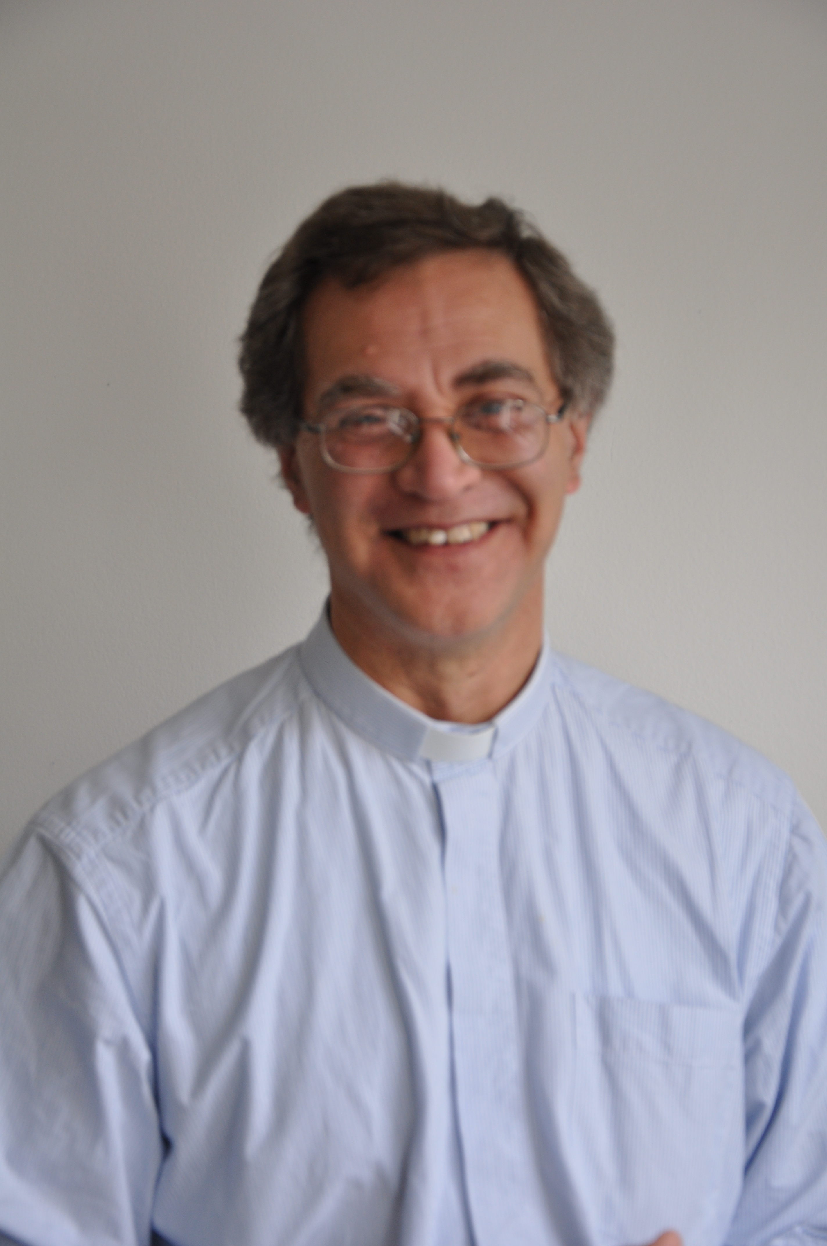
One of the first doctors to use radiology to diagnose diseases of the nervous system, Artur Schüller began his first systematic survey of the skull just a few years after X-rays were first discovered. Here, Andrew Schuller, a distant cousin, describes his extraordinary academic and personal journey which led to his recognition as the “Father of Neuroradiology”.

Artur Schüller (hereafter Arthur Schuller) was born in the Moravian city of Brunn (now Brno in the Czech Republic) in 1874. Most of the Schullers in Brunn were involved in the textile industry but Arthur’s father, Jonas, was an ENT specialist. Arthur did well at the German-language secondary school and went on to enrol in the medical school of the University of Vienna, which at that time had an excellent international reputation. He graduated in 1899 sub auspiciis Imperatoris, a rare title only awarded by the Emperor to students who had scored perfect marks in all their school and university exams. This entitled him to select his post-graduate mentors and Arthur chose Wagner-Jauregg and Kraft-Ebbing, a combination that matched his interest in both anatomy and psychiatry.  They sent him off to Berlin for 6 months in 1901 where he worked with Munk, Oppenheim and Krause who taught him about experimental physiology, clinical neurology and the diagnosis and treatment of brain tumours. By the time Schuller returned to work at the Allgemeine Krankenhaus, Vienna had already taken up Roentgen’s 1896 discovery of X-rays and Arthur was soon working with Guido Holzknecht, leader of Vienna’s radiological research efforts.

Arthur Schuller

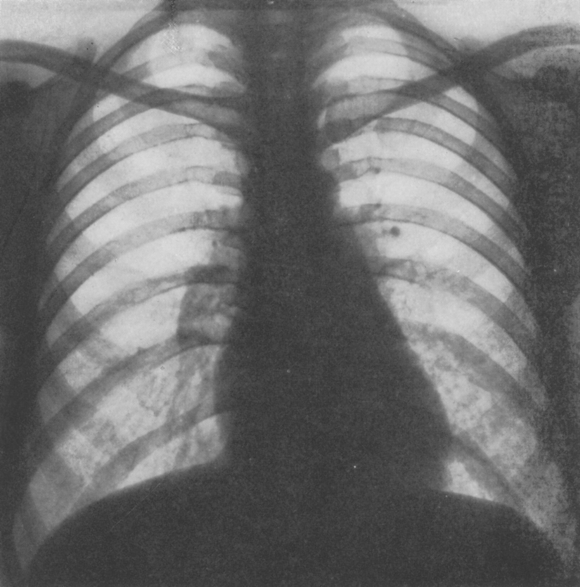
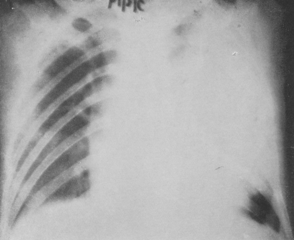
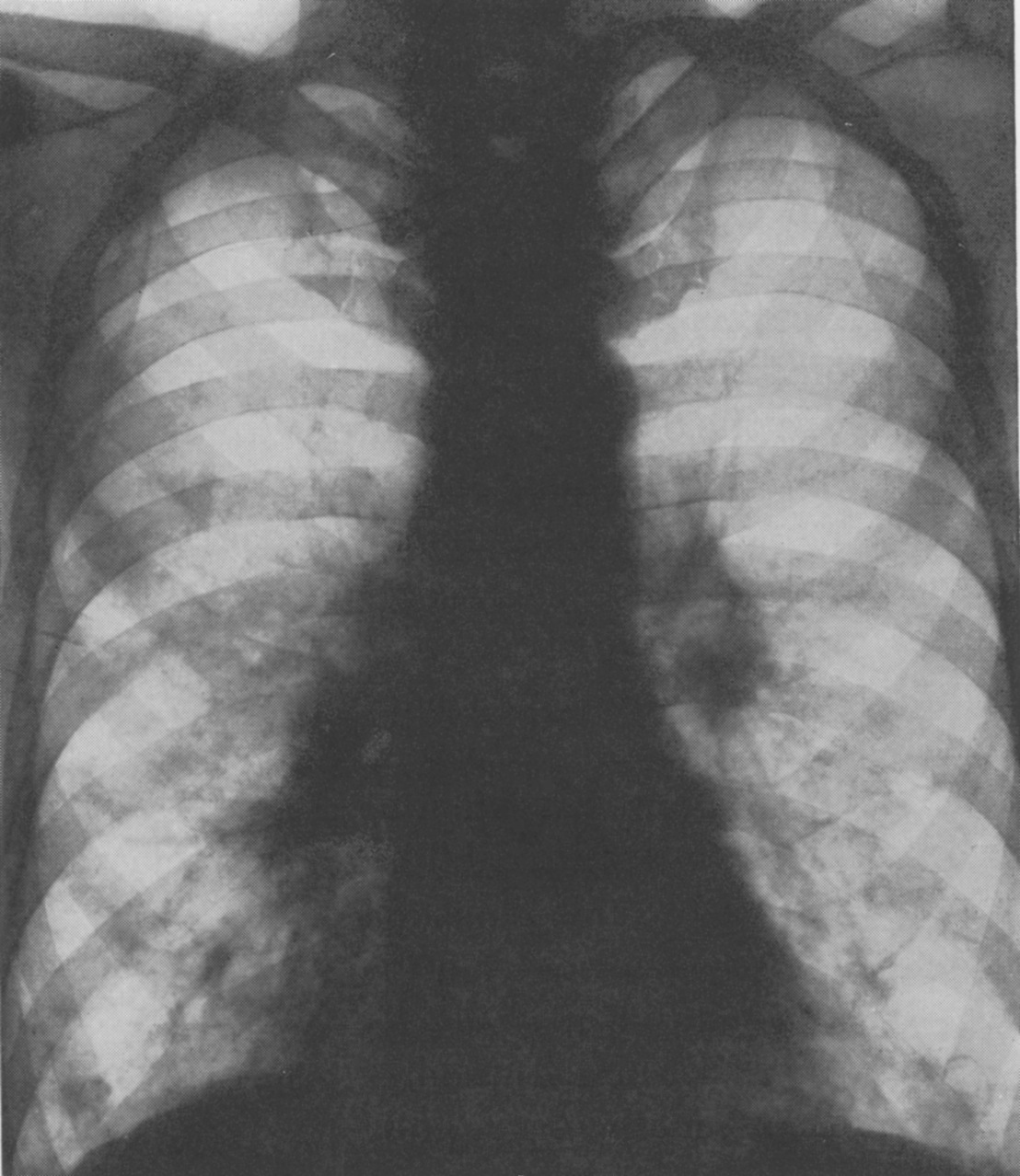
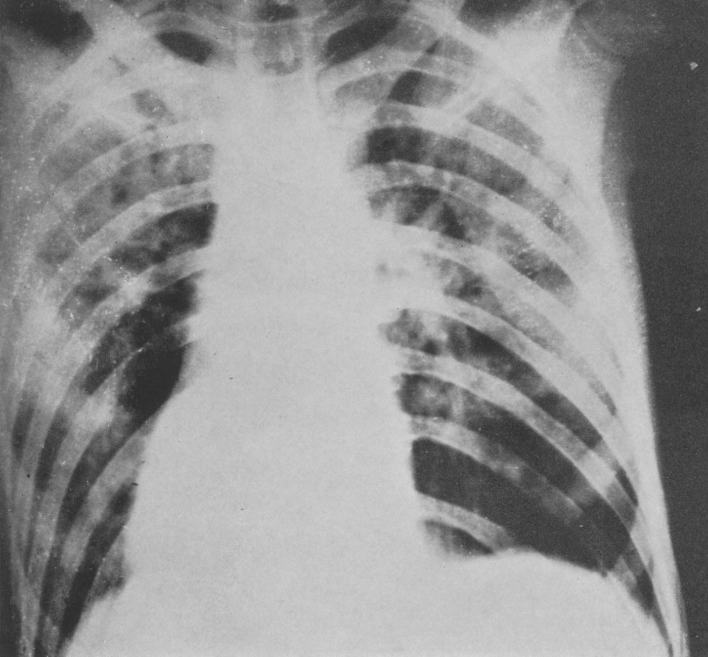
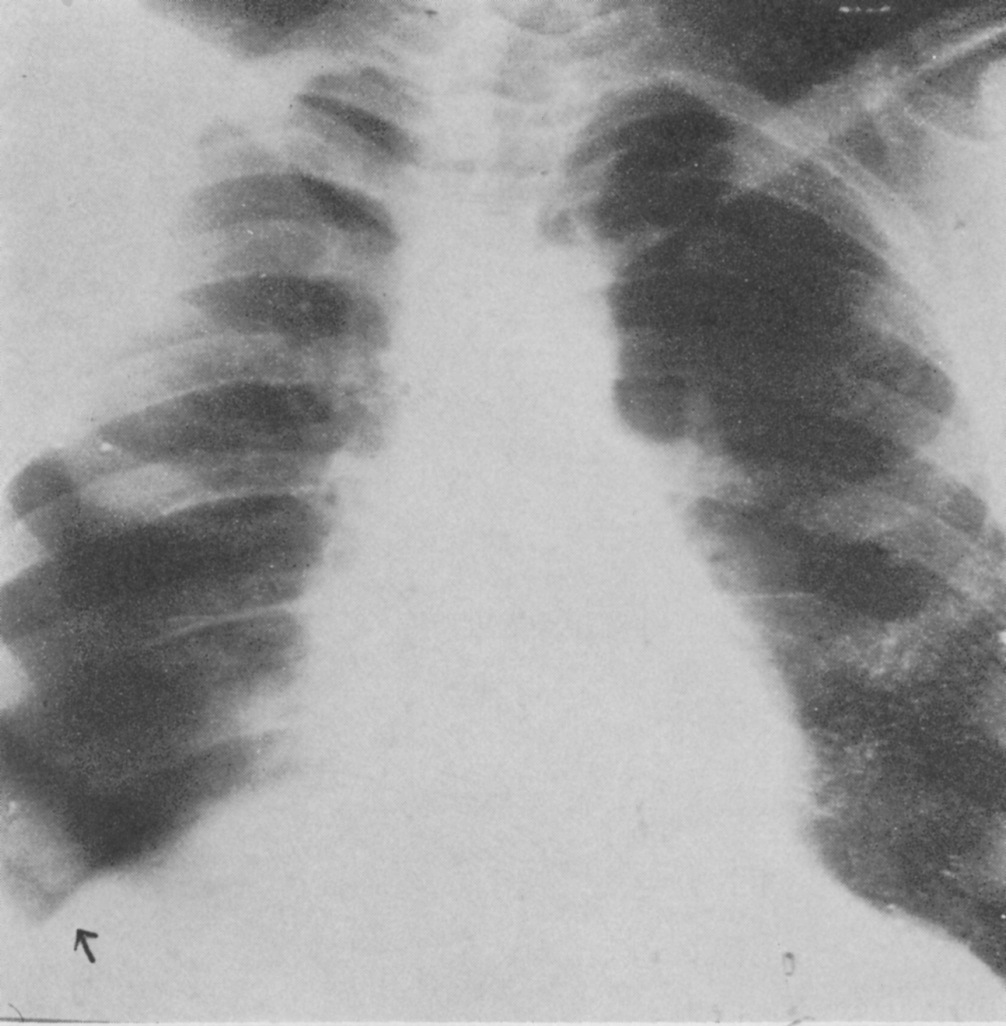
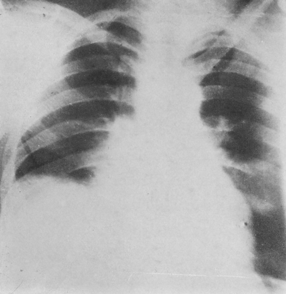
Schuller was the first to describe the role of X-ray in diagnosing diseases of the nervous system. Working with dried skulls from the museum and with live patients his painstaking analysis of countless X-rays enabled him to produce the first systematic survey of the radiology of the skull, which described both normal and pathologic anatomy. This book The Skull base on the Radiogram (Die Schädelbasis im Röntgenbilde: Archiv und Atlas der Normalen und Pathologische Anatomie) was published in 1905 and was followed in 1912 by Röntgen-Diagnostik der Erkrankungen des Kopfes (Röntgen Diagnosis of Diseases of the Head)  which encapsulated his extensive work on a range of topics and was eventually translated and published in 1918 by C V Mosby in America under the auspices of the US Army (Incidentally, during WW2 Schuller, by that time in Australia, wrote about battlefield head injuries and worked in a military rehabilitation hospital).

Schuller’s interests ranged widely. From 1904 he was Director of the Children’s Hospital where he had worked in both the Neurology and the Psychiatric Clinics. But he maintained his experimental lab work and practice at the Allgemeine Krankenhaus. By 1907 he not only passed his Habilitation (PhD) but was also awarded Dozent status which allowed him to teach courses at the university as well as privately; he kept an X-ray machine in his home. When he was made a University Professor in 1914 he was the youngest in the medical faculty. In addition to his contributions to neurosurgical procedures (transsphenoidal approach to the pituitary, antero-cordotomy and hydrocephalic drainage) Schüller is associated with three bone diseases: the Hand Schüller Christian syndrome, osteoporosis circumscripta and cephalohaematoma deformans. But it is in his foundational work in forming the discipline of Neuroradiology that his outstanding contribution lies.

The financial stringencies and the political volatility that followed the collapse of the Austro-Hungarian Empire in 1918 had a serious impact on the ability of the Vienna Medical School to maintain its position at the forefront of medical research. But the continuing presence of so many prominent medical scientists enabled it to retain its international reputation. Along with Wagner-Jauregg Schuller was instrumental in expanding the existing post-graduate courses which attracted students from round the world. The US even established The American Medical Society of Vienna to administer the flow of almost 12,000 American students who enrolled in these courses between 1921 and 1938. Schuller continued to write papers and teach but he also consolidated his international reputation by travelling the world to lecture at conferences and clinics in the UK, Europe, Latin America and the US, where he lectured at the universities of Chicago, Johns Hopkins and New York and the Mayo clinic. In teaching and travelling he established strong personal contacts that would stand him in good stead including both Harvey Cushing and Walter Dandy in the US, both pioneering neurosurgeons with an interest in radiology. In 1935, while attending the Second International Neurological Congress in London, he met Hugh Cairns, an Australian neurosurgeon, who invited him to come to Oxford. Perhaps the apex of Schuller’s career was the central role he played in the first international congress of neuroradiologists. This, the First Symposium Neuroradiologicum was held in Antwerp in July 1939; the 22nd symposium will be held in 2022.  There are some who assert that it was the Swede Lysholm who really established neuroradiology by developing contrast radiography, though it was Dandy who first wrote about ventriculography.

Schuller may not have pursued contrast radiography because of the lack of research facilities in interwar Vienna or because of the eclecticism and breadth of his intellectual interests but there is no doubt that his work was foundational for the routine radiography of the sella and its environs and the diagnosis of pituitary tumours. At the eighth Symposium Neuroradiologium in 1967 Bull and Fischgold declared “Without a shadow of doubt Arthur Schuller was the father of neuroradiology”. Schuller’s papers are still quoted in current literature and the Austrian Neuroradiological Society awards and annual Arthur Schüller Prize.

The Antwerp Symposium 1939

Schuller’s private life also flourished. In 1906 he had married Margarete Stiassni from a family of successful textile industrialists in Brunn. Arthur and Margarete were introduced at a post-opera supper party at the Sacher Hotel in Vienna where they shared their love of music. Arthur was a very competent violinist and played in the Vienna Medical School orchestra. In spite of the unsettled political situation, Vienna’s cultural life was still extraordinarily rich and the Schullers participated actively. They lived in a flat close to the university and the hospital and owned a house in Brunn and a weekend cottage by the Danube north of Vienna. They had a comfortable though not extravagant lifestyle. Indeed, the son of Arthur’s urologist cousin Hugo Schuller, who lived round the corner, reported that Arthur and Margarethe were somewhat parsimonious. Their two sons were born in 1908 and 1909.  It may be that dedication to Arthur’s profession and his travel schedule had some impact on his relationship with Franz and Hans. It seems that they were closer to their mother’s family in Brunn than to the other Schullers in Vienna. They spent at least some of their teenage years living in Brunn with Margarete’s mother, to whom they were devoted, and both decided to join the family business rather than go to university, with fateful consequences.

Arthur’s 60th birthday. Grandmother and sons are highlighted. Arthur Schuller is 5th from right and his wife Margarete is 6th from right.

The unstable political situation in Vienna deteriorated further in the late 1930s and after the Anschluss in March 1938 life for Jews became very difficult. Although Arthur and Margarete had been baptised as Roman Catholics in 1908 the National Socialist decrees categorized them as Jews. As such Arthur was only allowed to treat other Jews and in April 1938 he was officially “sent on holiday” from the university along with more than half of the members of the Medical Faculty, a purge that effectively set the Vienna Medical School back 60 years. The Nazi rampage through Vienna in November 1938 persuaded the Schullers that it was time to leave. Dandy had invited them to go to the US but Arthur was concerned about growing anti-semitism in academic medical circles there and decided on Australia. It seems that he was encouraged by two Australians who had attended his courses in Vienna and were now significant figures in Australian medical circles. Both John O’Sullivan and Sydney Sunderland were in Oxford in 1939 and to Oxford was where the Schullers fled when their Australian visas and their German Reich passports and exit papers arrived in early 1939. Arthur had followed up on Cairns’ invitation and in April was welcomed and attached to the labs of Le Gros Clark in Anatomy and Barclay at the Nuffield Institute for Medical Research.  While there Oxford Arthur wrote a paper on the sub-arachnoid cisterns and their demonstration using a positive contrast agent which was published in BJR in 1940, 13.(148):pps 127-29

The Symposium in Antwerp finished on 29 July, 1939 and in the first week of August the Schullers left Croydon airport on a KLM flight which took over a week and 30 stops to reach Darwin and then Brisbane and eventually on to Melbourne. Their arrival was noted in the press in the main Australian cities.

What the Australians had organised was a position at St Vincent’s Hospital in Melbourne run by the Sisters of Charity, which is where John O’Sullivan was based in the Radiology Department. Also at St Vincent’s was Frank Morgan the first specialist neurosurgeon in Australia. Schuller enjoyed spending time in both departments viewing and reporting on all head X-rays and attending Morgan’s ward rounds and operations. He was liked and respected by staff at all levels. Curiously, however, the Medical Board of Victoria did not recognise his University of Vienna qualifications and he was not formally permitted to practise in Victoria till 1946. He was lent rooms where he did see patients who were referred to him. He continued to write papers – his last was published in 1950 – and, although he declined to attend the second Symposium Neuroradiologicum in Rotterdam in 1949, he was elected Honorary President and the paper he submitted was given pride of place.

Dr Schuller and Dr Frank Morgan

In spite of his age Schuller’s attendance at St Vincent’s was constant, falling off only in his eighties. It was also stoical since on top of beginning to suffer from Parkinsons he had to bear the personal sorrow of family tragedy. Although they could have escaped, the Schullers’ two sons decided to stay in Brunn partly to care for their grandmother and partly to try to rescue the Stiassni family business. Both of them along with their grandmother and Hans’ wife and young daughter perished in Auschwitz in early 1943, though this news did not reach Australia till 1945. Arthur became increasingly depressed and even asked Morgan to perform a frontal lobotomy which Morgan refused to do. Arthur died in 1957 aged 83.

Margarete survived till 1971. She had taken to offering her services as a domestic help and, as such, worked for a number of Melbourne families. She cooked, ironed and looked after children. It is not clear why she did this. Since she left a substantial estate and her brother had continued to send monthly remittances from the USA financial need was probably not her main motivation. More likely she needed social company. She certainly embedded herself in some of the families for whom she worked. She was a devoted member of her local Catholic Church community.

Members of two of those families are featured in a 30 minute documentary film about the Schullers which is available for free viewing on YouTube at https://youtu.be/YhRLobn-Ubw

 Also featured is Dr Keith Henderson who, as a young neurosurgeon at St Vincents, worked with and befriended Arthur. Henderson wrote a biographical memoir of the Schullers entitled Arthur Schuller Founder of Neuroradiology: a Life on Two Continents which Hybrid Publishers in Melbourne have just, in February 2021, published posthumously *.  Henderson’s book contains substantially more detail about Schuller’s contributions to medical science than the film and it lists about half of the 300 papers he published.

*To order Arthur Schuller Founder of Neuroradiology: a Life on Two Continents  in the UK https://www.amazon.co.uk/s?k=Arthur+Schuller%3A+Founder+of+Neuroradiology&i=stripbooks&ref=nb_sb_noss

To order Arthur Schuller Founder of Neuroradiology: a Life on Two Continents  in Europe https://www.amazon.com/s?k=Arthur+Schuller%3A+Founder+of+Neuroradiology%3A+A+life+on+two+continents&i=stripbooks-intl-ship&ref=nb_sb_noss

About Andrew Schuller

Andrew Schuller

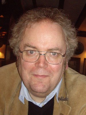
Andrew Schuller was born in and educated at Oxford. He worked for over 30 years for Oxford University Press in New York and Oxford. Now retired, he spends much of the year in Australia and continues to be engaged in some publishing projects as well as family history. Andrew’s grandfather was a first cousin of Arthur Schuller, though it is not known if they ever met. By a strange series of coincidences, Andrew became involved in helping Keith Henderson in the writing of his memoir. It was at the suggestion of Austrian historians who have been recording the career and fate of Jewish medical practitioners in Vienna that Andrew embarked on making the film. He regrets that he did not know about Arthur much earlier when it would have been possible to talk to more people who knew him in Austria, Oxford and Australia.

Adapting to a new way of treating

Dr Ben George

Over the last 18 months, GenesisCare has treated more than 170 patients on the UK’s first ViewRay MRIdian MR-linac and adopted SMART planning as a new way of working. Here, Ben George explains why this latest hypofractionated technique has proven to be one of the success stories of the COVID-19 era.

Stereotactic ablative radiotherapy (SABR) is growing in importance in the curative cancer pathway. Increasingly, it offers patients the opportunity to enjoy relatively long periods of disease control where previously they would have been considered for palliative treatments. During COVID-19, the scales have tipped even further in favour of hypofractionated techniques because protocols have been revised to limit the risk of patient infection. More recently, attention has turned to stereotactic ablative MR-guided adaptive radiotherapy (SMART) – the most exciting development in radiotherapy for years, with the potential to treat previously inaccessible targets.

GenesisCare has been the first in the UK to adopt SMART, installing the first ViewRay MRIdian MR-linac just over a year ago. Since then, we have treated over 170 patients, some of which are the most challenging in the world from a radiotherapy perspective, such as pancreatic, central lung and now renal cell carcinomas. MRIdian sits within our SABR offering, which is run by a specialist team of oncologists, physicists, dosimetrists, and radiographers. Over an intensive 18 months, we have adopted a completely new way of working and overcome the challenges of a pandemic to treat patients not just from across the UK, but also from around the world.

SMART explained

The MRIdian MR-linac combines a 0.35 T split superconducting magnet with a 6 MV linear accelerator. This gives it unique advantages over conventional external beam radiotherapy linear accelerators, which rely on kV cone-beam CT (CBCT) imaging, and enables an entirely new approach to treatment.

First, using MRI instead of CBCT provides superior soft-tissue visualisation. This increased imaging capability allows the treatment to be adapted at each fraction based on the daily position of the target and nearby organs at risk (OARs). This is in marked contrast to external beam treatment with CBCT, where anatomy captured in the CBCT is simply rigidly matched against a planning CT. This rigid registration is then used to calculate the movements required to shift the patient into the correct position for treatment.

Second, the MRIdian takes images continuously throughout the treatment period to not only monitor the patient position, but also turn the treatment beam on and off. This is carried out as the patient’s anatomy moves through the breathing cycle.

This combination of enhanced visualisation and real-time imaging adds a layer of certainty in the delivery of treatment.

  • The MRIdian on-table adaptive planning system generates a new, optimised treatment plan for each fraction. This accounts for these day-to-day anatomical variations when the patient is in the treatment position.
  • Treatment delivery is then automatically gated so that the dose is only delivered when the target is in the optimal position. The machine is able to monitor every intrafraction motion caused by breathing or organ-filling.

As a result of these factors, we can design plans which deliver a higher dose, more precisely than with conventional SABR. There is no need for invasive fiducial marker insertion and any uncertainty is removed. Moreover, we can reduce planning target volumes, remove internal target volumes, and minimise the amount of tissue irradiated.

SMART has led to a paradigm shift in how some cancers are treated. In particular, it can benefit cancers in areas where there is significant inter- or intrafraction motion of either the target or OARs. Across the global community, MR-linac centres are now treating novel indications, such as renal, central lung and hepatobiliary tumours, and achieving clinical outcomes not previously thought possible. It is not simply a case of improving on an existing treatment – for some tumour types, SMART is facilitating new referral patterns for patients who may not typically be eligible for radiotherapy.

Pancreatic cancer – a new way of treating

Pancreatic cancer is one such example and of all the tumour sites we are now treating at GenesisCare, this is undoubtedly the one that is breaking most ground, offering new hope for both clinicians and patients.

For decades, surgical resection and adjuvant chemotherapy and radiotherapy have been the cornerstones of primary and secondary hepatobiliary tumours and pancreatic cancer treatment. However, options are limited for many patients. Less than 20% are resectable at diagnosis and not all patients are fit enough for an operation or effective chemotherapy regimens. There is, however, emerging evidence of a dose-response relationship, proving that escalated radiation doses are associated with improved local control as well as overall survival in borderline resectable (BRPC) or locally advanced pancreatic cancer (LAPC). Conventional radiotherapy delivers a comparatively homogenous radiation dose to the target volume. In contrast, SABR treatments combine advanced image guidance systems, accurate dose delivery and hypofractionated regimes. This is to facilitate a deliberate heterogeneous dose distribution across the target. This means the radiation tolerances of surrounding OARs are respected, while the tumour receives a higher, ablative radiation dose. A number of SABR studies have yielded good results in the treatment of large hepatobiliary tumours, with 1-year local control exceeding 90% and acceptable toxicity. Furthermore, delivering these hypofractionated ablative doses of radiation over a shorter treatment schedule has the potential to reduce the burden of treatment on patients.

However, with conventional SABR this therapeutic approach is often limited by concerns regarding organ motion and the possibility of developing small bowel radiation toxicity. As a result, many patients are only being treated with systemic agents. This is a prime example of where the elements of SMART on an MR-linac can facilitate an effective radiation dose escalation, while still respecting the radiation tolerance of normal tissues and surrounding OARs. In fact, using an MR-linac, it has been possible to successfully increase the prescribed dose in patients with primary pancreatic cancer. The previous standard dose was 33 Gy in five fractions, but SMART enables us to escalate the prescribed up to 40 Gy or even 50 Gy in five fractions. At the time of writing, 30 patients have been treated on the MR-linac for pancreatic tumours at GenesisCare.

Compassionate Access

The significance of MR-linac as an innovation in cancer treatment can’t be understated and, although at GenesisCare we are offering it in a private setting, we are committed to sharing the benefits of this technology with the wider medical community. Patients with localised pancreatic cancer have variable access to precision radiotherapy in the UK. The n-SARS-CoV-2 pandemic has further disadvantaged this patient group by reducing the availability and safety of surgery and chemotherapy. Considering this, since 2020 GenesisCare in association with GenesisCare Foundation, UK charity, Pancreatic Cancer Research Fund, ViewRay and University of Oxford have been treating NHS patients with localised pancreatic cancer with SMART at no costThe programme, which is run through a partnership with the University of Oxford, is generating preliminary clinical and patient-reported outcome data on a UK cohort. This will inform the design of subsequent randomised clinical trials and help to embed SMART in UK oncology practice.

A new way of working

With any new technology, there comes a learning curve. MR-linac represents a significant change in working practices. It demands a style of inter-disciplinary working which challenges the norms.

In a standard radiotherapy workflow, a patient will receive a treatment planning CT one to two weeks before the start of treatment. During this time, several steps are carried out by a team of dosimetrists, physicists, doctors and radiographers to produce a treatment plan ready for the patient’s first fraction. These steps include contouring the treatment target and OARs and optimising the machine parameters to deliver the prescribed dose to the target while sparing critical structures. This is followed by reviewing the dose distribution, checking the planning process to ensure no errors have occurred and performing an independent dose calculation.

As part of the on-table adaptive workflow, the time taken for this process must be reduced from days to minutes. In order to achieve this, close inter-disciplinary working between the team is required. The need to undertake a number of complex tasks during each adaptive treatment also increases the time for each fraction to around one hour.

The MRIdian workflow involves a Clinical Oncologist on-site during treatment to oversee the daily adaption. To maintain a treatment schedule at GenesisCare, this has meant that clinicians had to be trained to contour all areas of anatomy, often working outside their main area of specialism. Equally challenging was the need to acquire skills in MRI interpretation, which for some specialities is not routinely used as a diagnostic modality. These were all skills that needed to be honed and validated before any patients could be treated on the MR-linac. In our case, we spent many hours learning with colleagues in MR-linac centres of excellence around the world. Twelve months later, we are experts in this field and have treated over 170 patients.

A body of evidence

There is a growing body of data as the global MR-linac community treats ever more and complex cases. We brought this international best practice to GenesisCare and have treated complex and challenging cases, including central lung, pancreas and reirradiation within our first year. We have many case studies available on our website genesiscare.com/mridian/case-studies. We already knew that the technology could deliver, but it was the confidence in our processes and the ability of our team to implement an adaptive workflow in a time-pressured environment, with a patient on the treatment table, which allowed us to embrace the opportunity that MR-linac presents in radiotherapy.

GenesisCare will install the second MR-linac in the UK in 2021. Through our MagNET programme, we are joining with NHS organisations to support education in the use of MR-guided radiotherapy. Enquiries to: James.Good@genesiscare.co.uk

Dr Ben George, Lead Physicist – MR Linac, GenesisCare

Ben is Lead Physicist – MR Linac at GenesisCare UK. He works as part of a multi-disciplinary team which has established a successful and world-leading SABR service delivering complex MR-guided adapted treatments. He has a PhD in Physics with a strong background in computer science, research and clinical computing. He has over ten years of experience as a Clinical Scientist specialising in radiotherapy in both the NHS and the private sector, and as a research scientist for the University of Oxford.

A podcast radiating positivity

Angela Young explains how the process of making a podcast helped not only others with a diagnosed brain tumour but gave comfort and support to herself as she embarked on a course of radiotherapy.

A brain tumour diagnosis, like all major events, can set in place a chain of emotions, among them anger, fear and denial. It can also make you adjust your priorities in life. I went through all this in 2015 when I discovered I had a Grade 1 benign posterior fossa meningioma. A resection at Addenbrooke’s Hospital in Cambridge was very successful, leaving only a 3mm residuum.

I had been having regular follow up scans, and in 2019, it was thought the growth was significant enough to consider radiotherapy. After the initial shock, I realised that, if successful, it would prevent the cells from growing again and remove the need for annual scans with the associated “scanxiety”. My decision to go ahead now rather than wait for symptoms to appear was influenced by the consultant radiologist Dr Sarah Jefferies who said the benefit of doing so now was that I was “young and fit”, a nice thing to hear at the age of 59.

As a journalist and podcast maker, I am used to getting to grips with a variety of subjects quickly in order to explain them to others. It dawned on me that if I could tell the story of my own treatment, it would give me a sense of control over a process in which one can easily feel helpless. It might also provide information and some light relief to other people going through something similar and their families. The radiotherapy process would be the same for people undergoing treatment for a variety of conditions, not just brain tumours, and so creating a podcast on this topic could reach and potentially help a large audience.

I am very optimistic by nature and I like to see the funny side of things. I believe that if you look closely, you can find humour in most situations. Consequently, I decided the title of the podcast should be “A Sense of Tumour”. I started recording everything that happened, whether by phone call (I had got all the kit I needed for doing this when lockdown started) or recording my own commentary during appointments and tests and arranging interviews, either face to face (with masks on) or via an audio recording platform.

People find podcasts in a variety of ways. One of those is to have a well-known personality or influencer or support group post about them. It helps if you can interview a celebrity or two who will do this. When I asked Victoria Derbyshire (via a mutual friend) if she would talk to me about documenting her very public battle against breast cancer, I had no idea she would later be taking part in the TV programme “I’m A Celebrity, Get Me Out Of Here!”. Victoria appeared in Episode 1 and set the interview bar quite high. Luckily, the Brain Tumour Charity had come on board by this stage and offered to put me in touch with TV presenter Nicki Chapman, who had had a matching meningioma to mine removed last year. She readily agreed to be interviewed and candidly shared the highs and lows she experienced when going through treatment herself. For the final episode, I thought I would chance my luck and ask to speak to Tony Iommi, lead guitarist and song writer with Black Sabbath. He had had radiotherapy a few years ago and embraced some alternative therapies which I wanted to hear about. To my delight, he was more than willing to talk.

The series was meant to inform as well as entertain so I spoke to the medical professionals whom I was meeting and also those at the cutting edge of research into treatment. I interviewed the Chair of Cancer Research UK, Sir Leszek Borysiewicz, about funding for brain tumours. I also had conversations with the “Distinguished Scientist” from Elekta, one of the companies which makes the linear accelerator machine (not a bad job title!) and to many people from the team at Addenbrooke’s, including a medical physicist and a research radiographer. I learned a lot and I hoped that sharing these conversations would also help listeners to understand some of the more complicated parts of the treatment and process more easily.

Bringing the podcast’s listeners on my journey was supposed to feel personal too. I recorded as much as I could at every stage, including the baseline neurological assessment. This is an IQ-style test carried out before the start of a course of radiotherapy to the brain so that if there is any concern about future cognitive function, there is a baseline against which to compare it. One part of the test included listing as many words as possible beginning with the letter F; you can imagine what came to mind! When that episode was released, listeners I came across would shout out words beginning with F to me.

All the way through the treatment I was thinking how I would represent things aurally, such as the MRI machine. These make a variety of loud noises but would wreck any recording device in the vicinity. When I managed to open my eyes under the thermoplastic mask which holds the head in place on the linear accelerator, part of the machine going over me looked like a spaceship. Friends and family had each contributed a song for my radiotherapy playlist; that day the song was Mr Blue Sky and it had got to the instrumental part, which made me think of a science fiction movie. I was working out how to recreate the impression for the podcast. Thinking about this during the session took my mind off what was going on.

By the time you read this, I will have finished the treatment and will be waiting for a scan to see how successful it has been. I am, of course, hoping for the best. I would also like to think that the podcast series has been useful to patients and their families, to radiotherapists, to manufacturers and anyone else involved in this fascinating process. I also hope that it inspires anyone looking for a positive and creative way of dealing with a diagnosis of any kind to take control of what they can, focus on something meaningful and use their good days to bring strength to others. After all, positivity radiates.

Listen to “A Sense of Tumour” here

About Angela Young

Angela Young founded Cambridge Podcasts in 2018 to help clients showcase their expertise and establish themselves as the go-to person in their field. She is a former BBC radio journalist who has worked as a reporter, producer, news reader and news editor. She has taught law and journalism at the BBC and media handling at the prestigious Institute for Management Development in Lausanne. She studied law at Cambridge as a mature student and has lived in the city for 28 years. www.cambridgepodcasts.co.uk info@cambridgepodcasts.co.uk

Key components to delivering successful regional imaging programmes

Jane Rendall examines what is needed to make large scale NHS imaging programmes work.

Jane Rendall

Region-wide NHS procurements for diagnostic imaging are becoming increasingly ambitious in their efforts to support faster diagnoses and better care for millions of patients. They are a means to reduce variation, to enhance equity, to change how healthcare professionals access, analyse and report, and they can facilitate new ways of working collaboratively across large geographies.

But what is the key to making these multi-trust projects successful and what lessons can be shared?

I’m fortunate to have worked alongside several NHS consortia that are doing some extremely impressive things. These are some of the key things, in my opinion, that can drive success.

1. Think multi-ology rather than radiology

As an early discipline to digitise, radiology has traditionally been the home for digital imaging in the NHS. But modern programmes are about realising a vision to bring imaging together from across the whole of healthcare. 

An integrated multi-ology approach to imaging means a much richer information resource to augment a report, and much more information at the fingertips of many more professionals, and indeed patients themselves.

Taking what has become known as an “enterprise approach” to imaging strategy isn’t a new idea, but it needs to be recognised and put into practice if large scale programmes being embarked on are to be successful.

2. Enterprise level responsibility and strategy

To move to an enterprise approach effectively, responsibility needs to be taken at the enterprise level. This might mean lifting imaging technology out of the radiology department and entering it as an IT or digital service.

There will probably be a range of internal funding and management complexities to be navigated in the process, but this will mean that imaging can sit at the heart of digital strategy and that information vital to patient care can flow more freely. 

It will also mean that radiology imaging system managers will not be burdened with an isolated task of managing data from multiple additional departments.

Sensitivity needs to be shown to people who might be concerned about losing niche functionality, and mechanisms put in place to maintain the tools they need.

A higher-level strategy that has the buy-in from chief executives, chief information officers, chief clinical information officers, finance directors and others, and that is clear on what organisations involved are going to achieve, must also be established.

The same “top brass” must also be willing to provide strategic sponsorship, leadership, and decision making at necessary points. Roadblocks will inevitably be encountered as large-scale programmes are conceived and executed. Strategic decisions will be continually needed along the way to overcome anticipated and unforeseen challenges, and those decisions cannot be taken in the ranks. When authority is provided from the top, a mandate can be given to allow the willing entrepreneurial people delivering great things on the ground to drive forward the initiative.

3. Managing complexity at a regional level

Elevating imaging to the enterprise level of a trust is one thing, but how can this be effectively done across an entire region? Some of the programmes I have worked with have as many as seven, eight, or even nine trusts involved.

One answer is to contract under one participating organisation which in turn has memorandums of understanding in place with all of the others. There are proven mechanisms that can work, but contracting is just one consideration in a programme that involves navigating everything from data sharing to the allocation of human resource.

That is why authority provided by strategic regional board level sponsors is key; if this is a priority for CEOs in a region, then it becomes a priority for others to work through some of these significant challenges.

But equally key is establishing appropriate governance structures, and putting the right people in place to manage delivery. Successful programmes that I have seen are driven by a type of “civil service”: people who do the job, who ensure strategic papers are passed through necessary boards and who can ensure the right things happen at the right time with the right people.

As a supplier, I think we must have faith in our customers and their ability to do this effectively, but again it comes back to having the right strategic objective with appropriate milestones, and a full understanding of how decisions affect different people throughout the organisations involved.

4. Finding the champions – and not just “yes people

Finding champions early is extremely important. For example, artificial intelligence is an area which is of huge interest. But even something as exciting as this will go nowhere without a champion who is really looking to push the idea within the customer organisation; someone who can talk to people and engage, energise and inspire them. Champions must have a level of power to drive the conversation and they need to have aligned goals.

Once the champions have been found, it’s important not to take them for granted and to show them appropriate recognition. This might mean helping to elevate their voice as an influencer or thought leader in the media, for example, or nominating them for awards for which they are deserving.

Successful programmes need more than “yes people” though. You also need champions who are prepared to challenge. That’s when you really start to have useful dialogue. You want people who spot the problems before they happen, people who know the operation and who know their business. We need the people who really understand technologies and what we can do outside of the norm, so that when a problem requires an unconventional solution, the art of the possible can become reality. As a supplier, that sometimes means knowing when to take an anti-sales approach and to bring the technical experts to the table.

5. Work with the exiting supplier

Exiting suppliers have a significant role to play in smooth transitions, especially for areas like data migration.

It can be challenging for a supplier that might be disappointed to be the outgoing party. But it is important for that supplier to consider long-term relationships and their reputation in the market.

Where we have exited in the past, we have made sure we have delivered a professional, timely, responsive approach to that exit. It is a long-term game and a small world in NHS technology. In 5 or 10 years we might well be back there.

6. Patience

Programmes of this scale do not happen overnight. That’s not to say that given the momentum building in the NHS for regional diagnostic approaches that accelerated approaches cannot be developed. But with so many parties involved, things can take longer than a lot of people might predict. Be patient and maintain your determination.

7. An open mind

Customers can often benefit from approaching procurements with a truly open mind. Sometimes people can have a preconceived idea of what the solution needs to look like before they go to market. Sometimes getting real value from the market means allowing competitive suppliers to come up with the innovative solutions to meet the requirement.

Some suppliers in the market have learned lessons from similar projects that have gone before, lessons that they are often more than happy to share. And they may have visibility of new technology coming down the line that the contracting organisation isn’t necessarily aware of.

Intelligent customers know their strategy, they know their organisational needs, the requirements of their people and they know their business inside out. But they also know that there are opportunities to be educated at times. There might be ways of solving problems that they hadn’t previously considered. And in some major procurements we have seen the end solution looking very different to the original requirement.

8. Vision

Establishing and communicating a central vision is crucial throughout all of the above. This must be bought into from the most senior CEO to the user on the ground. Given the pressures faced in NHS diagnostics, and the opportunities for transforming patient care that come from a regional approach to imaging, the vision behind many regional initiatives is extremely powerful.

About Jane Rendall

Sectra UK’s Managing Director, Jane Rendall, joined Sectra in 2010. Located in our UK and Ireland headquarters in Hertfordshire, Jane has a strong clinical background and is on a mission to drive innovation through collaboration and evolve healthcare IT to more cost efficient and adaptable platforms to improve the effectiveness of patient care across the UK and Ireland.

The Life of Brain

What do tiny bees and dead salmon have to do with the history of MRI? This post by Dr David Higgins and Dr Matthew Clemence explores how the flexibility of MRI lends itself to important applications outside of medicine and examines how the use of functional MRI has more recently brought us much closer to real scientific observation of the brain.

MRI is a rapidly evolving imaging modality, and the history of MRI has always been intertwined with research. Its flexibility lends itself to fascinating applications outside of medicine; even bees, and fish.

In neurobiology, honeybees are a common model for analysing underlying neural mechanisms because of their simply structured nervous system. By investigating the bee brain’s anatomy, correlations between anatomy and function can be studied. See Haddad et al 2004 for MR images of tiny bee brains. One early project looked to see if there were any magnetic structures inside the bee – which MRI was uniquely sensitive to – that might help bees navigate. Bees have been studied by MRI surprising often, perhaps because their behaviour is extraordinary, emerging as it does from such apparently simple creatures. MRI has helped our understanding of this important crop animal (Tomanek et al, 1996).

Looking at the function of intact, living neuroanatomy has been a dream for students of the human mind for centuries. Phrenology (from Ancient Greek φρήν (phrēn) “mind”, and λόγος (logos) “knowledge”), which supposed that the lumps and bumps on the skull reflected personality traits, was developed in 1796 and remained influential up until the 1840s. While, in a marked understatement from Wikipedia, “the methodological rigor of phrenology was doubtful even for the standards of its time”, the underlying concept that that the brain is the organ of the mind – and that certain brain areas have localised, specific functions – is based in fact. Technology developments in electroencephalography, near-infrared spectroscopy and most recently functional MRI have brought us much closer to real scientific observation of the brain.

Which brings us to a dead salmon…

Functional MRI (fMRI) was a revolutionary technique used to identify highly localised changes in blood flow resulting from differential load on regions of the brain. John (Jack) Belliveau at Massachusetts General Hospital showed in a key Science paper that these changes could be measured with MRI (Belliveau et al, 1991) in conjunction with a Gadolinium contrast agent, but it was Seiji Ogawa who demonstrated a practical, non-invasive method with the University of Minnesota (Ogawa et al, 1990). For the first time, scientists had access to detailed 3D anatomical images of the brain in action, through a safe technique that could be repeatedly used on volunteers. Its ease of use, combined with wide availability, allowed the technique to be adopted rapidly in psychology and neuroscience, often in the hands of the non-MRI specialist. Some of the early studies exploring this new ability to “read minds” often drew overly broad conclusions from badly designed experiments. This almost relegated fMRI into a category of “modern phrenology”.

The dead salmon experiment showed how, with naive experimental design and data analysis, fMRI could give convincing results on a dead Atlantic salmon (Bennett et al, 2010) and was a salutary lesson to would be fMRI researchers to improve their methodology (Lyon, 2017).

Now, once again fMRI is being used to tease out our inner thoughts, whether to attempt to detect lying for legal purposes or read letters directly from the visual cortex. This could have a dramatic impact in patients with “locked in syndrome” through the development of brain computer interfaces (Sorger, 2010).

fMRI has also found an unusual application in neuromarketing: the application of neuroimaging methods to product marketing, to more effectively “match products with people”. Companies can incorporate use of fMRI in the design process of a product, as well as in assessing the effectiveness of an advertising campaign, even if the “product” is a political candidate. “Political marketing is aimed at selling an existing candidate but, with more foresight, can also be used to “design” a better candidate” (Ariely & Berns, 2010). Imaging our brains may reveal what we really think (or how we’re likely to vote), even if we can’t fully articulate our preferences yet.

If all this sounds hard to believe, perhaps a look at our brain scans could help you decide whether to believe us. “The relative reduction in prefrontal grey matter relative to white may also predispose to a general antisocial disinhibited tendency which, coupled with increased white matter, results in excessive lying.” (Yang et al, 2005).

Just check the study passes the dead salmon test!

To see how Philips can help you in neuroscience visit:

https://www.philips.co.uk/healthcare/resources/landing/neuro-mr

Explore the Philips MR image quality in the Body Map at:
https://www.mriclinicalcasemap.philips.com/

To learn about the latest Philips MR innovations, please visit: https://www.philips.co.uk/healthcare/solutions/magnetic-resonance

References

Ariely D & Berns G. Neuromarketing: the hope and hype of neuroimaging in business. Nat Rev Neurosci 2010;11:284–292. doi:10.1038/nrn2795

Belliveau JW et al. Functional mapping of the human visual cortex by magnetic resonance imaging. Science 1991;254:716-719. doi:10.1126/science.1948051

Bennett et al 2009. Neural correlates of interspecies perspective taking in the post-mortem Atlantic salmon: An argument for proper multiple comparisons correction. J Serendipitous Unexpected Results 2010;1:1–5. See https://www.nature.com/articles/nj7420-437a

Haddad D et al. NMR imaging of the honeybee brain. J Insect Sci 2004;4:7. doi:10.1093/jis/4.1.7

Lyon L. Dead salmon and voodoo correlations: should we be sceptical about functional MRI? Brain 2017;140;e53. doi:10.1093/brain/awx180

Ogawa S et al. Brain magnetic resonance imaging with contrast dependent on blood oxygenation. Proc Natl Acad Sci U S A. 1990;87:9868-9872. doi:10.1073/pnas.87.24.9868

Sorger et al. A real-time fMRI-based spelling device immediately enabling robust motor-independent communication. Curr Biol 2012;22:1333-8. doi:10.1016/j.cub.2012.05.022

Tomanek B et al. Magnetic resonance microscopy of internal structure of drone and queen honey bees, Journal of Apicultural Research, 1996;35:3-9. doi:10.1080/00218839.1996.11100907

Wikipedia contributors. “Phrenology.” Wikipedia, accessed 16 Oct. 2020. Yang Y et al. Prefrontal white matter in pathological liars. Br J Psychiatry 2005;187:320-325. doi:10.1192/bjp.187.4.320

Dr David Higgins and Dr Matthew Clemence are Senior Scientists at Philips, part of the UK&I MR Clinical Science team and the wider global Philips Clinical Science group. They provide: MR physics support; advanced teaching on the functions of the MR system; prototype pulse sequence deployment and monitoring; novel pulse sequence development advice; guidance for novel image reconstruction and analysis projects; advice for novel interfacing novel hardware.

The five-step guide to AI adoption in clinical practice

Jeroen van Duffelen proposes a five-step programme for adoption of artificial intelligence in clinical practice.

Adoption of medical imaging AI is about getting your hospital or screening programme ready to implement the right solution for a clinical need. Running into speed bumps along the way is common for early adopters. How do you define the needs, budget, and outcomes? Which boxes should you check when selecting vendors? How do you manage internal stakeholders? The adoption curve is steep. Luckily, you don’t have to climb it alone.

Drawing from our experience deploying AI in clinical practice and lung cancer screening, I’ve designed a five-step guide to streamlined adoption. If you’re looking to adopt artificial intelligence but don’t know where to start, these actionable tips and advice will see you through. For the video breakdown of the steps, watch this presentation from ECR 2020.

1. Consider

Where do you start working with AI? First, look away from all the solutions out there, and focus on your organisation. Bring together all the stakeholders into a project team that includes the sponsor, if applicable, IT and legal representatives. Involving them from the beginning will expedite the process.

Start with defining the challenge you are looking to solve, or the specific clinical question that is relevant to your workflow. Some hospitals are looking to experiment with the technology, while others aim to solve a particular issue. Over the past years, I have seen the latter getting more out of AI, which is why my advice is to start from a clinical challenge.

When considering this challenge, make sure to already determine your expected outcomes. When is the adoption a success? Are you aiming to have an AI solution in use? Should it apply to a certain patient population, or yield specific results like time or cost savings?

Also, although it may seem early, this is also the stage to organise a budget dedicated to the AI solution. The size of this budget should relate to the cost or time savings a solution is expected to bring. Both the amount secured and its source will impact the next steps. For example, it will guide you to look for PhD researchers versus seeking a vendor that offers a mature solution.

2. Evaluate

The AI in healthcare space is widely populated; a Google search or a look at the list of vendors at the RSNA can confirm that. To weigh the existing options for your scope, do your (desk) research using this high-level checklist for each solution:

How was the AI solution validated?

It is important that the claim that the AI solution has validated covers the use case you identified in the previous step. Take the time to understand if the manufacturer has done studies confirming this claim.

How does it integrate into the workflow?

Try to get a feel of the amount of effort needed to add an AI system into your workflow. A good practice is to start with an AI solution that is easy to integrate with the current workflow and IT infrastructure. Workflow integration is of utmost importance for the radiologist; in this article, we explained why that is and how it works.

What regulations does the solution fulfill for use in clinical practice?

Commercialising medical devices requires a CE Mark in the EU and an FDA clearance in the US. Note that local regulations may apply to different countries. Again, pay attention to which claim is covered by the acquired certification.

3. Choose

By this stage, you should have narrowed your search down to a few vendors. This is the moment to go in-depth into the workflow and test if a specific solution is a good fit from both a clinical and a technical standpoint. A well-integrated AI system should not create hurdles for physicians, such as requiring them to leave their workstation to upload studies. It should further blend within the existing IT infrastructure.

There are two checks that are vital to make the right choice:

Validate the accuracy

Legitimate vendors would have done a study and can provide a clinical background for accuracy. To know if the solution is good at performing the defined task for your organisation, ask questions about the datasets used to develop and test the AI solution.

There are three datasets required to build an AI model: a training dataset, a validation dataset, and a test dataset.

The test dataset is the most relevant to look at because it is what the accuracy is based on. The performance on this dataset should be applicable to your hospital, with its specific protocols, type or number of scanners, and patients. To achieve this, the test set must cover the patient population your organisation serves (e.g. types of patients, comorbidities distribution, etc.). Thus, inquire about the specifics of the test set and the performance of the AI model on this dataset.

Secondly, you may want to know what the size of the training dataset is and how it was labelled. Both quantity and quality are important to train an accurate AI model. Labeling the data should be done by experienced radiologists, preferably with multiple readers per study.

Check the regulatory compliance

In Europe, medical device classification is divided up between risk Class I, Class IIa/b, or Class III. If looking for a solution for clinical practice, be wary of Class I medical devices. The new Medical Device Regulation, which will come into force in May 2021, will require many AI products currently classified as Class I devices to update their classification. For instance, software that supports diagnostic decisions should fall under Class II at a minimum. For more guidance on the new regulation, read our recent expert piece.

Apart from the regulatory approval, check if the vendor also has a quality management certification (e.g. ISO 13485). Reviewing the data processing policy and the cybersecurity measures in place will further help you understand if the AI company is going the extra mile in regard to safety.

A bonus tip for the choosing stage: do a reference check. Ask other organizations how they are working with the AI solution you have chosen. You may get the insights you need to make the final decision.

4. Approve

Approving the chosen solution internally requires the involvement of and coordination between IT and PACS administrators, procurement officers, physicians, often also privacy departments and legal officers. If you have a project team in place since the first step, you should be well on track.

To move forward and avoid delays, assign an internal AI champion responsible for driving the project. This may be an executive sponsor, a budget holder, or a department manager. One of my learnings from past deployments is that the risk of failure is high without a person fulfilling this role. What I have further learned as vendors is the importance of empowering the AI champion, by providing the necessary information and documentation in a timely manner.

Furthermore, make sure end users are trained to use the new medical device. If they don’t benefit from it, the impact of the AI solution will be limited. Additionally, setting up a feedback mechanism with the AI vendor from the get-go will help improve the AI product.

5. Deploy (& evaluate)

All the paperwork is signed – well done! To make the deployment work, create a clear project plan, including actions, timelines, and owners. Depending on the type of deployment – on-premise or cloud-based – different actions will be needed. As outcomes, set the deployment and acceptance dates, make agreements on the service levels, fixes, and upgrades, and discuss post-market surveillance.

The initial or trial phase of using the AI solution should show if it answers the problem you were trying to solve. It is a good moment to revisit step one and start evaluating the results to decide if you will continue using the solution.

A common question I get at this stage is: “Do I need to do a full clinical study?” The answer fully depends on the purpose of using the product. It is necessary for research, but not for other use cases. What matters is validating that the AI solution is adding value to your clinicians and their patients.

Make it better

AI adoption does not end with deployment. Service and maintenance are essential, and their quality often a differentiating factor between AI vendors. The implementation process usually acts as a good test for the AI companies fulfilling their promises and being prompt when handling requests.

Beyond these five steps, you and your organisation play a role in improving the chosen AI solution through valuable feedback and feature suggestions. The collaboration between humans and software allows us to achieve much more than humans would on their own. If done right, it can be transformative for patients.

Are you ready to start the AI journey? Get in touch!

Jeroen van Duffelen, COO & Co-Founder

Jeroen van Duffelen is COO and co-founder of Aidence. Jeroen’s entrepreneurial spirit led him to teaching himself software engineering and starting his own company commercialising an online education platform. He then tried his hand in the US startup ecosystem where he joined a rapidly scaling cloud company. Jeroen returned to Amsterdam where he ran a high-tech incubator for academic research institutes, it is here Jeroen first got his taste for applying AI to healthcare.

Is artificial intelligence the key to effective and sustainable lung cancer screening?

Lizzie Barclay doctor

Dr Lizzie Barclay explores how artificial intelligence can influence lung cancer screening.

Radiology as the starting point

Imaging plays a fundamental role in lung cancer screening programmes. So, when it comes to improving technology to support the programmes, the radiology department is a good place to start.

The goal of screening is to pick up early cancers which can be treated and potentially cured, therefore improving patient outcomes (as outlined in the NHSE long term plan). Low dose CT has been shown to provide sufficient image quality for detection of early disease, whilst minimising radiation dose in asymptomatic individuals. Thoracic radiology expertise is required to determine which lung nodules may be malignant and therefore require invasive investigation, and which are likely benign and can be monitored with intermittent imaging. Appropriate follow-up recommendation helps avoid unnecessary invasive procedures, such as biopsies, and minimise patient anxiety, which are important measures of the efficacy of lung cancer screening programmes.

End to end lung cancer screening involves input from many healthcare professionals, and intelligent computer systems across specialities would benefit multidisciplinary teamwork. Thus, beyond image analysis, there are many opportunities for technology to add further support for effective and sustainable screening programmes. For instance, it could aid in the optimisation of image acquisition, access to imaging reports and relevant clinical details, tracking patient follow up, or in communication between patients and GPs.

Where AI-based image analysis makes a difference

Reading and reporting CT scans is time-consuming, and within a workforce which is already under strain, introducing a new CT-screening programme seems like a tall order. AI-driven solutions can support radiologists and contribute to successful lung cancer screening by bringing improvements in three areas:

  1. Performance

Computer intelligence can increase the performance and productivity of CT reporting, freeing up time for radiologists to spend on clinical decision making and complex cases. Specifically, AI software is well-suited for precise:

  • Detection of elusive lung nodules, and differentiation of subtle changes
  • Automatic volume measurements, to help determine the appropriate frequency of monitoring (e.g. stable vs growing nodule, according to the BTS guidelines).

What further distinguishes computers from humans is the absolute consistency in their high performance, without being impacted by common external stressors to which a radiologist would be exposed (e.g. time-pressure, workload and interruptions).

  1. (E)quality

Having a ‘second pair of eyes’ looking at the scan can increase the confidence of the radiologist in their own assessment. Additionally, making the AI-driven, accurate measurements available regardless of the level of expertise of the reporting radiologist could not only benefit quality assurance, but also equality within the radiology department. The use of AI would reduce the need for all scans to be reported by the most experienced thoracic radiologists with interest in early lung cancer detection, and instead facilitate spreading the workload across the workforce.

Another use case concerns quality assurance when outsourcing to teleradiology companies. AI-based image analysis can improve consistency of reporting, drive the recommended terminology use, and, essential for lung cancer screening, ensure access to relevant prior imaging for comparison and change assessment over time.

  1. Efficiency (via integration)

An intelligent computer system should not slow down reporting turnaround times, but improve efficiency, as well as quality, to ultimately minimize time to diagnosis (for example, the NHSE long term plan introduces a 28-days standard from referral to diagnosis or rule out).

Older CAD technology was often described as ‘clunky’ – requiring images to be uploaded to separate systems for analysis. Additional manual steps between image acquisition and the radiology report make the process time consuming, and often require radiology support staff to manage the workflow. It is important to consider allocative and technical efficiency which play important roles in the evaluation of screening programmes, and their impact on healthcare systems.

An AI-driven image analysis software which is fully-integrated in the radiologist’s pre-existing workflow can provide automatic results, without needing additional departmental resources. An additional benefit of fully-integrated AI solutions is that their use is not restricted by time or place, therefore supporting flexible and remote working. In the context of the COVID-19 pandemic, it’s been encouraging to see the increase in remote reporting, whilst maintaining a functioning department, in many hospital trusts. Going forward, it will be interesting to see whether radiologists will have the option to continue to work remotely where possible.

Valuing input from healthcare professionals

New lung cancer screening programmes will be monitored regularly to evaluate their effectiveness and determine areas for review. Commitment from all parties to work together will facilitate optimisation of the pathway to achieve better patient outcomes and positive impacts on healthcare systems.

In our experience, close collaboration between medtech and healthcare professionals is important for learning lessons along the way. Understanding radiologists’ needs helps tech teams develop a clinically valuable tool.

For example, Aidence’s interactive lung nodule reporting tool, Veye Reporting, was designed based on the needs of radiologists involved in reporting lung screening scans. From our conversations with them, we understood that following the detailed and complex reporting protocols in lung cancer screening programmes make for labour-intensive, repetitive tasks.

Veye reporting

To help them produce reports that follow the standardised NHSE proforma and facilitate audit for quality assurance, we added Veye Reporting as a feature to Veye Chest, focusing on making it easy-to-use and efficient. With this tool, the radiologists further have control over which nodules to include in the report, different sharing options, and the choice to add incidental findings.

What’s next?

Cancer services have been impacted by the COVID-19 health emergency. In the UK, screening has been paused and planning to (re-) start at the end of 2020 or beginning of 2021. Talks of introducing screening are ongoing in various European countries, as are concerns of catching up with the backlog of screening scans.

The British Society of Thoracic Imaging and the Royal College of Radiologists released these considerations for optimum lung cancer screening roll-out over the next five years. Their statement below is a reminder of why it is worth overcoming challenges and leveraging technology to make screening programmes a success:

BSTI_RCR statement

Dr Lizzie Barclay, Medical Director

Dr Lizzie Barclay’s areas of interest are thoracic radiology and medicine, innovation, and improving patient outcomes and healthcare professionals’ wellbeing.

Lizzie is originally from Manchester, UK. After graduating from the University of Leeds Medical School (MBChB), and Barts and the London School of Medicine (BSc sports & exercise medicine), Lizzie spent four years working as a doctor in Manchester and Liverpool NHS Trusts, including two years in Clinical Radiology. She has presented her work on lung cancer imaging at national/international conferences, and recently contributed to Lung Cancer Europe’s “Early Diagnosis and Screening” event at the EU Parliament in Brussels.

Homepage

You may be interested in the BIR Lung Cancer Imaging: Update for the not-so-new normalon 11 September 2020. This will be available for members in the BIR online learning libraryafter the live virtual event.


“The Radiology Reset Button – overcoming the normalcy bias”

Fodi KyriakosFodi Kyriakos explores how the COVID-19 pandemic could be the catalyst for change in radiology and encourages our community to grasp the opportunity to “seize the moment”  and plan for recovery.

At the beginning of 2020, if someone had told radiology leaders that all NHS outstanding reporting backlogs would be reduced to virtually zero by May, I’m sure they would have looked at you in disbelief and asked what sorcery had been involved, but this situation is exactly where we find ourselves today.


Normalcy Bias – Noun [edit]

normalcy bias (plural normalcy biases)

The phenomenon of disbelieving one’s situation when faced with grave and imminent danger and/or catastrophe. As in over focusing on the actual phenomenon instead of taking evasive action, a state of paralysis.

Historical challenges

In the past, it has often taken lots of effort to either invoke or accept change of any kind in radiology and for those managing services, there’s also been a certain amount of risk associated with putting your head above the parapet or being a trailblazer. It has been sometimes easier to follow the well-trodden path rather than to create a new one. Workloads and budgetary constraints have also been a disabler, restricting decision making to the ‘here and now’. This has resulted in failing, or in most cases, not being able to foresee or plan for events that have never happened before, such as an event like a pandemic crisis. Psychology refers to this state of being as normalcy bias. For those who are not familiar with the term, you will certainly be aware of its connotations and radiology now finds itself at this cross-roads.

Ever since the introduction of digital radiography and PACS, NHS radiology reporting backlogs have been a contentious issue among experts, and a recurring feature in the mainstream media! Often being highlighted (and with some justification) in relation to areas such as missed cancer diagnosis, where even the slightest of delays can have a significant bearing on the overall outcome.

Serious backlogs

The extent to which backlogs were a serious issue in the UK was further exacerbated by various Care Quality Commission (CQC)  inspections, which raised concerns regarding reporting backlogs that resulted in delayed or missed diagnosis of conditions that may have otherwise been picked up.

By the end of February 2020, the situation of backlogs was as much an issue as at any time before. Insufficient reporting capacity had led to a build-up of outstanding reports, which in turn meant that outsourcing was at its highest ever levels and growing pressures to meet new deadlines, such as the cancer pathway targets, were increasingly exposing the lack of options available to resolve the problem.

So, you would have been excused if you thought that a crisis such as the COVID-19 pandemic would simply exacerbate the reporting challenges facing radiology. However, this has not been the case. Instead, we have witnessed radiology’s own “clear the decks” exercise, where in fact the complete opposite situation has occurred, resulting in backlogs across the UK being virtually eliminated. Who would have thought that the worst crisis to hit the country (and the world) in 75 years would be a catalyst for NHS radiology departments to press the reset button?

Reset image

Of course, we recognise the superficial nature of this situation. During the pandemic, practically all routine referral activity came to a grinding halt, which allowed radiology to concentrate on COVID-19 and Emergency Department (ED) patients. Chest X-rays and CTs were identified as two of the key diagnostic tools for the virus, but the volumes were manageable. Accident and Emergency footfall was reduced to almost 50% of its usual figures, so reporters were practically able to deliver a ‘Hot-Reporting’ examination for every patient requiring imaging. Something which ED and Intensive care unit (ICU) consultants have grown quickly accustomed to.

During this time, radiology was also still required to work to critical staffing levels, so radiographers and radiologists were covering 24/7 rotas, but due to the lack of activity outside of portable X-ray scanning in ICU, many staff were not being utilised. So, while this enabled the catch up in radiology reporting to take place, what we witnessed was the ‘ying and yang’ of radiology. On the one hand, integral to the continuity of a patient’s pathway and critical to defining an outcome – AND on the other hand, completely dependent on throughput from referrers to maintain activity levels.

Seizing the moment!

So what happens next? Well, in a world where we can guarantee almost nothing, in this situation, we can guarantee that radiology will remain the centre point for the recovery phase of the pandemic, but with the added challenge of complying to ‘social distancing’ and ‘equipment cleaning’ guidelines, how do we manage the continuation of treating COVID-19 patients, while reintroducing ‘business as usual’ and ‘deferred’ patients whose treatment has been delayed?

The “Reset Button” has enabled something else to happen. For the first time, there is now some headspace to plan for the recovery phase and for the next phase at least, there is now funding available to support the recovery. So how do we avoid going back to where we were before the pandemic? How do we seize the moment?

Time to make the changes!

Albert Einstein once famously said: “We can’t solve problems by using the same kind of thinking we used when we created them.” This quote has never been more poignant in the present day and while the pressure to manage change will be at its highest, this is the right time to make these changes happen! With the benefit of ‘The Reset Button’, if we can learn from the past and apply new ways of working moving forward, we can avoid falling into the trap of the normalcy bias and witness the radiology reset button offering a new, efficient and more streamlined radiology department moving forward.

Everything you wanted to know about radiology but were afraid to ask…

On Wednesday 17 June, a live event organised by InHealth, in partnership with The British Institute of Radiology and the Society of Radiographers is taking place, titled: “The Radiology reset button has been pressed”. The aim is to tackle these challenges and support radiology managers as they enter the recovery phase. It will bring together senior figures from radiology and within healthcare to offer insights, opinions and advice on how we can approach this coming period and use what positives we have experienced during the pandemic to create service improvements throughout radiology.

There will be opportunities for radiology managers, clinical leads, radiographers and radiologists to put their questions to the speakers in the panel discussions after their presentations.

REGISTER FOR THE RADIOLOGY RESET BUTTON HAS BEEN PRESSED HERE

(The event is free for all)
About Fodi Kyriakos

Mr Fodi Kyriakos is a former director of RIG Healthcare and founder of RIG Reporting,
the UK’s first provider of external radiographer reporting services. In 2016 he joined The InHealth Group following its acquisition of RIG Reporting and is now the Head of Reporting across the Group. His service specialises in delivering plain film reporting solutions and is the only provider to offer both on-site and telereporting services.
Fodi has over 22 years experience in workforce and staffing solutions and 17 years working exclusive within Imaging and Oncology. He is a member of the Institute of Healthcare Managers and a regular contributor of professional development events across radiology.


The flu epidemic of yesteryear: the role of radiology in 1918–20

adrian thomas

Dr Adrian Thomas

100 years ago the UK was facing a fast-moving outbreak of epidemic influenza pneumonia, known as the “Spanish Flu”.

Radiology played an important part in diagnosis, although the crisis was without the scientific knowledge, strategic management and communications we have today. Here, Dr Adrian Thomas explores the six patterns of infection in this unpredictable and powerful disease.


Radiology is playing a central role in the diagnosis of COVID-19 today, and 100 years ago was also playing an important role in the diagnosis and characterisation of the outbreak of epidemic influenza pneumonia of 1918–1920. A combination of fluoroscopy and radiography was then used, with the occasional utilisation of stereoscopy. The greatest pointer to a diagnosis of epidemic influenza pneumonia in a given patient was the presence of the epidemic, although there were some specific features to indicate the diagnosis. The etiological cause of influenza was not known at the time, being first discovered in pigs by Richard Shope in 1931.

Spanish flu

The epidemic of 1918 far exceeded previous ones in its intensity. It had a high mortality in young adults with the very young and very old being comparatively immune. The associated pneumonia was particularly virulent. In the case of the troopship The Olympic (sister ship of The Titanic) there were 5,951 soldiers on board. Initially there were 571 cases of acute respiratory disease, but within 3 weeks there were 1,668 cases. Of these, 32% had pneumonia, of which 59% died. In any locality the duration of the epidemic was from between 6–8 weeks, and approximately 40% of the population was affected (Osler, 1930).

Six patterns of infection were identified, with correlation of clinical, radiological and post-mortem findings (Sante, 1930., Shanks, et al. 1938). Dr Leroy Sante, the pioneer radiologist from St Louis, described epidemic influenza pneumonia as “the most lawless of the chest infections.” Abscess formation was seen frequently, and was commonly of the small and multiple type. Radiological changes were seen developing day by day, and clinical resolution needed at least six–eight weeks since there had commonly been lung destruction and healing by fibrosis needed to occur.

The patterns were:

Type 1: Peribronchial invasion with infiltrates that enlarge and become confluent forming small areas of consolidation (figures 1 & 2, below). This was not confined to one lobe, but could appear in all lobes as a true bronchopneumonia. This was similar in appearance to ordinary bronchopneumonia.

1Type 1, Influenza bronchopneumonia

Figure 1

2Type 1, Influenza bronchopneumonia_Viewed as from behind

Figure 2

Type 2: Peribronchial invasion with infiltrates that enlarge and become confluent to form solidification of an entire lobe (figure 3, below). The changes remained confined to a single lobe. It was viewed as a true bronchopneumonia but with a lobar distribution (“pseudolobar pneumonia”). Different lobes may be invaded one after another. The pseudolobar pattern was the commonest type, and could resolve without further spread. The presence of previous isolated infiltrates would distinguish this type from common lobar pneumonia. There was a tendency to break down with extensive cavitation.

3Type 2, or pseudo-lobular

Figure 3

Type 3: This starts as blotchy infiltrates that coalesced to form a general haziness over a part of a lung, suggesting a haematogenous origin (figures 4 and 5, below). At post-mortem this was found to be an atypical lobular pneumonia, a “diffuse pneumonitis”, that was so commonly seen during the influenza outbreak. It resembled the streptococcal (septic) pneumonia that was often seen in association with septicaemia when there was no epidemic. The spread was rapid, and the prognosis was poor. Death commonly occurred within the week.

4Type 3, resembling streptococcal (septic) pneumonia

Figure 4

5Type 3, resembling streptococcal (septic) pneumonia

Figure 5

Type 4: A type starting in the hilum and spreading rapidly into the periphery, the so-called “critical pneumonia” (figure 6, below). This was attended with a high mortality. Post-mortem showed a purulent and haemorrhagic infiltration around the larger bronchi. There was often marked cyanosis.

6Type 4, the so-called “critical pneumonia

Figure 6

Type 5: This started in the dependent part of the lungs, with continuous upwards spread (figures 7a and b, below). This was an atypical lobular pneumonia, there was no associated pleural fluid, and it was usually fatal. Initial infection in the costo-phrenic angle spread within 24 hours to involve the lower lung, and death occurred within 48 hours. Clinical features included extreme prostration, high temperature, and delirium. This pattern with rapidly advancing consolidation was seldom seen in other conditions.

7a Type 5. This started in the dependent part of the lungs

Figure 7a

7b Type 5. A film taken 12 hours after 7a

Figure 7b

Type 6. A true lobar pneumonia was only seen rarely.
The prognosis of epidemic influenza pneumonia was difficult to determine. So, as an example, a patient who was resolving would suddenly have changes extend into the other lung and then die. Another patient with successive involvement of all lobes could recover completely. A patient with only minor lung involvement might die, and another with extensive consolidation would recover completely.

Radiologists continue to be in the front line in the treatment of infectious diseases, and although our modalities are now more advanced than a century ago, their contributions remain essential. It is also noteworthy that the simple CXR also remains central.

Figures:

1. Type 1, Influenza bronchopneumonia. Image seen as a positive.

2. Type 1, Influenza bronchopneumonia. Peribronchial clusters of infiltration, with no relation to lobar architecture. Viewed as from behind.

3. Type 2, or pseudo-lobular.

4. Type 3, resembling streptococcal (septic) pneumonia. Image seen as a positive.

5. Type 3, resembling streptococcal (septic) pneumonia. Blotchy ill-defined infiltrates which coalesce to form a general haziness. Viewed as from behind.

6. Type 4, the so-called “critical pneumonia.”

7a. Type 5. This started in the dependent part of the lungs, and this early film shows consolidation in the costophrenic angle (black arrow).

7b. Type 5. A film taken 12 hours after 7a. The lower right lung is consolidated, and the patient died 12 hours later. Post mortem showed a solid lung with no effusion.
Readings:

Osler, William. (1930) The Principles and Practice of Medicine. 11th Edition, Thomas McCrae (Ed.). London: D Appleton.

Sante, Leroy. (1930) The Chest, Roentgenologically Considered. New York: Paul B Hoeber.

Shanks, S Cochrane., Kerley, Peter., Twining, Edward W. (Eds). (1938) A Textbook of X-ray Diagnosis by British Authors. London: H. K. Lewis.


Dr Adrian Thomas FRCP FRCR FBIR, BIR Honorary Historian

About Dr Adrian Thomas

Dr Adrian Thomas is a semi-retired radiologist and a visiting professor at Canterbury Christ Church University. He has been President of the Radiology Section of the Royal Society of Medicine, and of the British Society for the History of Medicine. He is the Honorary Historian to the British Institute of Radiology. He has had a long-term interest in role development in radiography, and teaches postgraduate radiographers.

Adrian has written extensively on the history of radiology writing many papers, books and articles. He has, with a colleague, written a biography of the first female radiologist and female hospital physicist: Adrian Thomas and Francis Duck: Edith and Florence Stoney, Sisters in Radiology (Springer Biographies) Springer; 1st ed. 2019 edition (1 July 2019).

© Thomas / 2020

Bringing together Science, Faith and Cancer Care

Slide2

The Revd. Canon Dr. Mike Kirby, Chair of the BIR Oncology and Radiotherapy Special Interest Group, has a wealth of experience as a senior radiotherapy physicist, working on national guidance, developing clinical practice and teaching radiography students. As if this doesn’t keep him busy enough he has also taken on the role of Canon Scientist at Liverpool Cathedral where he is working to encourage dialogue and discussion about science and faith. Here he explains what the role involves.

I began work in the UK’s National Health Service more than 30 years ago, as a Radiotherapy Physicist at the Christie Hospital, Manchester UK.  Alongside my routine clinical work, my main research interest was in electronic portal imaging and portal dosimetry.  I then helped set up Rosemere Cancer Centre in Preston, UK from 1996 as deputy Head of Radiotherapy Physics and Consultant Clinical Scientist there.  During that time I contributed to and edited national guidance documents such as IPEM Reports 92, 93 and 94 and the multidisciplinary work, ‘On-target’.

My work moved back to the Christie in 2007 and as Head of Radiotherapy Physics and Consultant Clinical Scientist for the Satellite Centres, I helped to lead their development in Oldham and Salford as part of the Christie Network. My research and development work has primarily focused on electronic portal imaging, developing clinical practice and equipment development.

Mike Kirby4

More recently my focus has been on teaching and learning for radiotherapy education as a lecturer (Radiotherapy Physics), especially using VERT, for Radiotherapy programmes in the School of Health Sciences, Liverpool University; but always with a focus on the wider picture of radiotherapy development having served on both IPEM and BIR committees throughout my whole professional career.


Alongside my scientific work, I am a priest in the Church of England; having trained and studied at Westcott House and the Universities of Cambridge and Cumbria, I hold graduate and postgraduate degrees in Theology.

Mike Kirby

My ministry has mainly been in the Cathedrals of Blackburn, Chester, and Liverpool (Anglican) where I was Cathedral Chaplain.  I have recently (Feb 2020) become a Residentiary Canon of Liverpool Cathedral, with the title of Canon Scientist the primary aim of which is to encourage dialogue and discussion about science and faith.

 I am a member of the Society of Ordained Scientists and have given numerous talks on Science and Faith to schools, colleges, churches and other institutions.  These have included organising lecture series with world renowned speakers at Blackburn (2016) and Chester (2018) cathedrals; a third series was delivered at Liverpool Cathedral in May 2019, and a fourth series is planned for May 2020.

My role is to consider all sciences (physical, clinical, social) in ecumenical and multi-faith environments.  So I will look to work with initiatives already developing in other Christian traditions, other faiths and secular organisations to discuss current challenges, such as climate change, medical ethics, health initiatives and information for cancer, dementia and mental health issues etc..

Mike Kirby2.jpg

My work will be part of the clear faith objectives of the cathedral as a place of encounter for everyone, through events and initiatives within the cathedral, but also beyond.  This will include services focusing on health issues and pastoral challenges (such as bereavement and loss); events engaging with science, its wonders and challenges; fostering further relationships with local and wider communities on science and healthcare education, and with academic and scientific institutions too; encouraging scientific and ethical engagement with schools and colleges, as I have done so previously in both Chester and Blackburn dioceses.

I will be encouraging Christians and Christian leaders to understand science and engage with it more, alongside other national projects such as the recently announced ECLAS (Engaging Christian Leaders in an Age of Science) project of Durham and York universities and the Church of England.  As a self-supporting minister (one whose paid employment is outside of the church), I will also look to encourage and highlight the tireless work of many others who already do this within the diocese and the wider national church.

Within all of this, I have always seen my vocation as being one within God’s service, for all people, with my work for cancer patients being right at the heart of it.

006

If you have any questions for Mike, you can send him an email at sigs@bir.org.uk

Mike is the co-author of the international student textbook on On-treatment Verification Imaging: a Study Guide for IGRT, through CRC press/Taylor and Francis with Kerrie-Anne Calder. They are both contributors to the updated UK national guidance on IGRT due out in 2020.

Mike, with the support of the SIG, has helped to organise a range of events for radiographers, physicists, dosimetrists, radiologists and oncologists. See the full programme here